Midline sagittal T1WI.

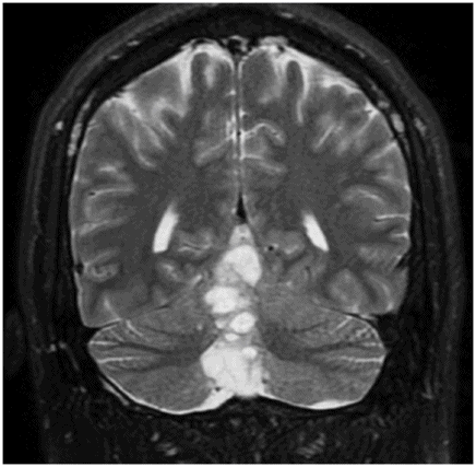
Coronal T2WI through the cerebellum.
Rosette-Forming Glioneuronal Tumor
Primary Diagnosis
Rosette-forming glioneuronal tumor
Differential Diagnoses
Dysembryoplastic neuroepithelial tumor (DNET)
Pilocytic astrocytoma
Medulloblastoma
Ependymoma
Imaging Findings
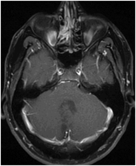
Fig. 83.1: Sagittal T1WI showed multiple, small, rounded and oval-shaped hypointense lesions in the cerebellar vermis with minimal mass effect. Fig. 83.2: Coronal T2WI showed multiple hyperintense nodules in the cerebellar vermis, with no adjacent vasogenic edema. Fig. 83.3: Axial T1 postcontrast image showed multicystic appearance of the lesion, without any enhancement. It is to be noted that the cystic lesions extend beyond the margin of the fourth ventricle.
Discussion
In a young adult patient, the presence of a midline vermian multicystic tumor without enhancement is suggestive of rosette-forming glioneuronal tumor (RGNT).
Although RGNT was initially described as DNET of the posterior fossa, true posterior fossa DNET is exceedingly rare. Pilocytic astrocytoma is typically a cystic tumor, with an intensely enhancing solid component. Medulloblastoma typically demonstrates diffusion restriction and is a solid or predominantly solid tumor (with few cysts), rather than a purely cystic tumor. Ependymoma of the fourth ventricle arises from the ependymal lining of the ventricle and is an intraventricular tumor. Extraventricular posterior fossa ependymoma is extremely rare, and thus excluded.
Rosette-forming glioneuronal tumor of the fourth ventricle (RGNT) is a recently described mixed tumor that expresses neuronal and glial differentiation with or without a ganglion component. They are rare, benign (WHO grade I), slow-growing, indolent tumors composed of distinct neurocytic rosettes or perivascular pseudorosettes and an astrocytic component. Typically, they are found in young female patients (30 ± 12.8 years – 2× more common than in males).
Recognized as a distinct entity in 2002, as opposed to DNET of the cerebellum, RGNT was thought to occur exclusively in the fourth ventricle and the adjacent parenchyma. It is now known that this tumor can occur in other CNS locations including the pineal region, optic nerve, and spinal cord. In some patients, these tumors are asymptomatic and are discovered incidentally. When symptomatic, patients commonly complain of headache, vomiting, ataxia, vertigo, neck pain, and cranial nerve palsies. Owing to the typical location (fourth ventricle), patients may show obstructive hydrocephalus.
On CT imaging, RGNTs are predominantly hypodense tumors, with few areas of calcifications. On MRI imaging, RGNTs are well-defined solid-cystic masses which are T1 hypointense and T2 hyperintense with variable degrees of enhancement (ring or heterogeneous) but little mass effect or surrounding edema. In one-quarter of reported cases, these lesions show calcification. These lesions are typically in the midline, and primarily involve the floor of the fourth ventricle with dorsal invasion of the vermis and ventral invasion of the brainstem either directly or as a satellite lesion. Although satellite lesions are common, the exact mechanism of satellitosis is not understood. However, satellitosis may be a clue to the diagnosis. From the fourth ventricle, RGNTs may extend to the cerebral aqueduct. The tumor can be completely solid or completely cystic, or may be mixed. Focal enhancement may be seen and can be nodular, ring-like, or linear. Typically, RGNT does not involve the lateral recesses of the fourth ventricle. Intratumoral hemorrhage may be seen.
On pathology, RGNT specimens are known to demonstrate dual histologic characteristics composed of neurocytic and astrocytic/glial components, hypothetically originating from the periventricular germinal matrix. The neurocytic components are formed by small cells with hyperchromatic nuclei forming rosettes disposed around an eosinophilic core stained with synaptophysin and absent vessels. The organization of the astrocytic/glial component with eosinophilic processes resembles pilocytic astrocytomas, with positive immunostaining for glial fibrillary acid protein (GFAP) and S-100 protein.
Generally, surgery is the treatment modality of choice for RGNT. It has been documented that total or subtotal surgical resection results in satisfactory tumor treatment, with excellent outcome and minor morbidity.
Stay updated, free articles. Join our Telegram channel

Full access? Get Clinical Tree


