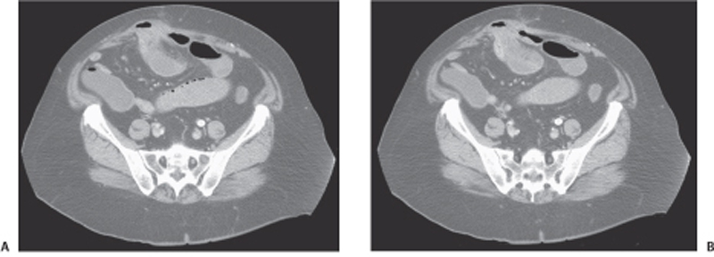
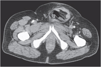
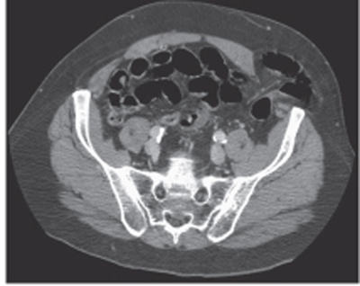
CASE 83 A 57-year-old man presents with abdominal pain and discomfort. Physical examination reveals a palpable mass in the anterior abdominal wall, in the midabdomen. Fig. 83.1 (A,B) Axial CT image show small bowel loops herniating through the abdominal wall and surrounded by a rim of soft tissue density with stranding, consistent with mild inflammation. There is mild distention of the small bowel proximally to the herniated loops. Axial computed tomography (CT) images (Figs. 83.1) show small bowel loops herniating through the abdominal wall and surrounded by a rim of soft tissue density with stranding, consistent with mild inflammation. There is mild distention of the small bowel proximally to the herniated loops. Abdominal wall hernia Fig. 83.3 Axial contrast-enhanced CT image shows a left ventral hernia containing small bowel loops and mesentery. A hernia is commonly defined as the protrusion of any viscus from its proper cavity into a saclike structure formed by the same membrane with which the cavity is usually lined. Different types of hernias have been described according to their anatomical distribution, the most frequent in both men and women being inguinal hernias (direct Fig. 83.2 and indirect). Others include
Clinical Presentation
Radiologic Findings
Diagnosis
Differential Diagnosis
Discussion
Background
Related posts:


Stay updated, free articles. Join our Telegram channel

Full access? Get Clinical Tree


