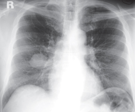
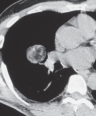
CASE 83 40-year-old man with hemoptysis PA (Fig. 83.1) chest radiograph demonstrates a 3 cm mass with well-defined lobular borders in the right mid-lung. Unenhanced chest CT (mediastinal window) (Fig. 83.2) shows a well-defined polylobular middle lobe mass with intrinsic fat and soft-tissue attenuation as well as a small focus of punctate calcification. Hamartoma None Pulmonary hamartoma is a benign pulmonary neoplasm composed of mesenchymal tissues, including cartilage, fat, connective tissue, and smooth muscle. These tissues are found in varying proportions. Entrapped respiratory epithelium may also be found within the lesion. Hamartomas account for approximately 8% of lung neoplasms, are considered the most common benign tumor of the lung, and represent approximately 77% of all benign lung neoplasms. Fig. 83.1 Fig. 83.2 Pulmonary hamartoma is thought to arise from peribronchial mesenchymal tissues, but its etiology remains unknown.
 Clinical Presentation
 Radiologic Findings
 Diagnosis
 Differential Diagnosis
 Discussion
Background
Etiology
Clinical Findings
Related posts:


Stay updated, free articles. Join our Telegram channel

Full access? Get Clinical Tree


