Case 84
Indication: Small lump in the right breast.
History: Unremarkable.
Risk profile: No increased risk.
Age: 39 years.
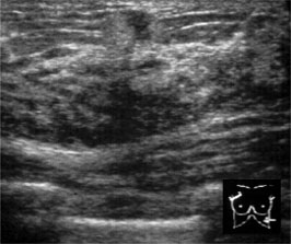
Fig. 84.1 Sonography.
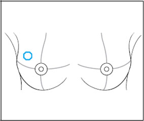
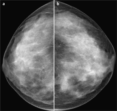
Fig. 84.2 a,b Digital mammography, CC view
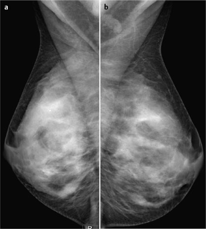
Fig. 84.3 a,b Digital mammography, MLO view.
Clinical Findings
A small resistance with characteristics of a gland measuring 5 mm in the upper outer quadrant of the right breast. Generally nodular parenchymal texture.
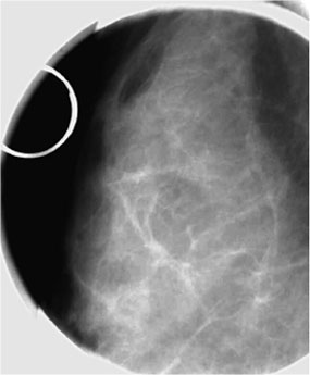
Fig. 84.4 Spot compression of the upper outer quadrant of the right breast with marker at the site of the palpable mass.

|
Please characterize ultrasound, mammography, and MRI findings.
What is your preliminary diagnosis?
What are your next steps? |


