Case 84
 Clinical Presentation
A 38-year-old woman with flank pain. She underwent mitral valve replacement 3 months previously.
 Imaging Findings
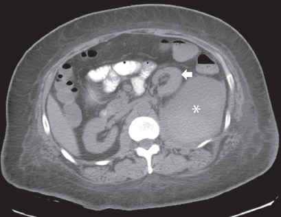
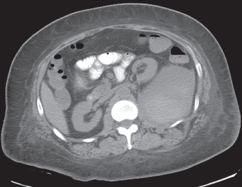
Noncontrast axial computed tomography image at the level of the kidneys. The left kidney (arrow) is displaced anteriorly by a large lesion (asterisk) in the left psoas muscle. The lesion shows heterogeneous attenuation, with areas of high and low attenuation. No calcification is seen.
 Differential Diagnosis
• Psoas hematoma: Bleeding into the psoas muscle appears as a collection with high attenuation when it is acute and more fluid attenuation with the passage of time. A history of anticoagulation is usual.
• Psoas abscess:
Stay updated, free articles. Join our Telegram channel

Full access? Get Clinical Tree


