Clinical Findings | right 1 | right 1 |
Ultrasound | right 1 | left 1 |
Mammography | left 1 | left 5 |
BI-RADS Total | right 1 | left 5 |
This case demonstrates the relevant images of a woman within a screening situation.
Ultrasound
As expected in the present case, ultrasound showed no unusual findings, including in targeted examination of the area behind the nipple and in the inner quadrants of the left breast.
Mammography
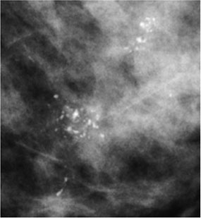
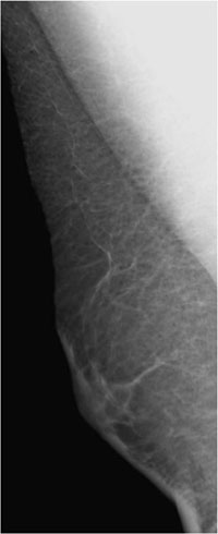
Markedly dense parenchyma, ACR type 4. Under these limiting conditions, no density or circumscribed lesion was visible. There was no architectural distortion. Mammograms depicted several clustered distributions of polymorphous calcifications in the lower inner quadrant of the left breast. In combination, the distribution of these calcification clusters was segmental. BI-RADS left 5. PGMI is not defined for unilateral mammography.
Procedure
Histological analysis of the microcalcifications with stereotactic vacuum biopsy. Calcifications were found in 9 of the 12 specimens (Fig. 85.3).
Histology
Extensive ductal carcinoma in situ (high-grade with necroses) with signs of microinvasion of the breast.
Fig. 85.3 Specimen radiography.
Histology
Minimally invasive ductal carcinoma. Axillary lymph node status normal.
DC pT1 mic, pNO (0/4)sn, VNPI 7-8 points.
Stay updated, free articles. Join our Telegram channel

Full access? Get Clinical Tree


