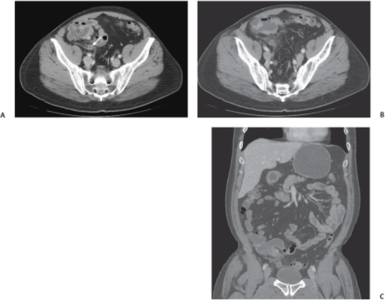
CASE 86 A 24-year-old man presents with right lower quadrant pain, fever, and nausea. Fig. 86.1 (A,B) Axial contrast-enhanced CT images demonstrate appendiceal enlargement and wall thickening (arrow) with stranding in the periappendicular fat (mesoappendix). (C) Coronal reformatted image shows the thickened appendix along with periappendiceal inflammatory changes. Axial and coronal contrast-enhanced computed tomography (CT) images (Fig. 86.1) demonstrate appendiceal enlargement and wall thickening with mild stranding in the periappendicular fat (meso-appendix). A calcified appendicolith is noticed within the appendix. These findings are consistent with acute appendicitis. Acute appendicitis Appendicitis represents the most common cause of right lower quadrant pain, usually occurring in young patients of both genders with a slightly higher male predominance. This disease requires prompt surgical treatment to avoid complications; for this reason, radiologists should be able to recognize this entity as promptly as possible. Appendicitis usually presents with right lower quadrant pain and tenderness, fever, anorexia, nausea, and vomiting. However, the clinical presentation can be protean and mimic other pathologies. The variable position of the appendix, the degree of inflammation, and the timing of onset can all contribute to a difficult clinical diagnosis. Laboratory tests typically show a leukocytosis with left shift.
Clinical Presentation

Radiologic Findings
Diagnosis
Differential Diagnosis
Discussion
Background
Clinical Findings
Complications
Stay updated, free articles. Join our Telegram channel

Full access? Get Clinical Tree


