Case 87
Indication: Monitoring following treatment for breast cancer.
History: Breast cancer 5 years previously. Breast conservation therapy.
Risk profile: Increased by earlier instance of breast cancer.
Age: 47 years.
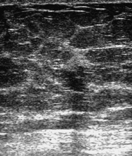
Fig. 87.1 Sonography.
Clinical Findings
Normal scar after breast conservation ther apy (BCT). No palpable mass.
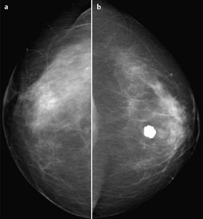
Fig. 87.2a,b Digital mammography, CC view.
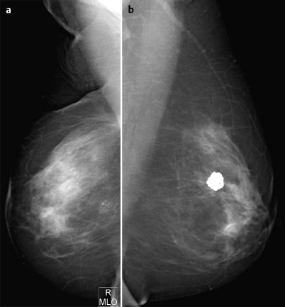
Fig. 87.3a,b Digital Mammography, MLO view.
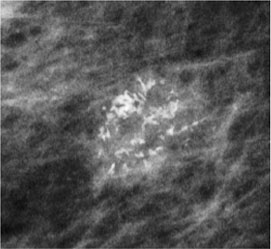
Fig. 87.4 Magnification view of the right breast close to the chest wall.

|
Please characterize ultrasound, mammography, and MRI findings.
What is your preliminary diagnosis?
What are your next steps? |


