Case 87
 Clinical Presentation
A 67-year-old woman with a history of cervical cancer.
 Imaging Findings
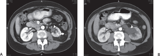
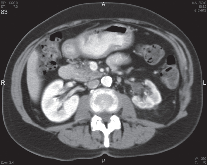
(A) Contrast-enhanced computed tomography (CT) image in the region of the kidneys. The left kidney (arrow) is significantly smaller than the right because of parenchymal atrophy. The outline of the unilateral small kidney is smooth. There is a mild delay in perfusion. The collecting system shows mild dilatation (arrowhead). The contralateral kidney is normal. (B) Contrast-enhanced CT image of the abdomen at the same level 6 months earlier confirms left hydronephrosis (arrowhead) in the past. This was produced by a malignant pelvic lymph node (not shown) and was treated with a ureteric stent in the interval between the two scans.
 Differential Diagnosis
• Postobstructive atrophy: Unilateral smooth atrophy of a kidney with residual dilatation of the collecting system is characteristic. A demonstration of hydronephrosis in the past confirms the diagnosis. The contralateral kidney becomes hypertrophic if the event took place when the patient was younger than 40 years of age.
• Long-standing unilateral renal vascular disease:
Stay updated, free articles. Join our Telegram channel

Full access? Get Clinical Tree


