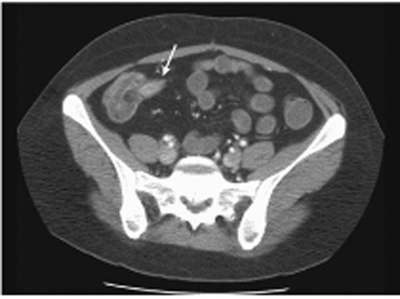
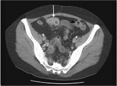
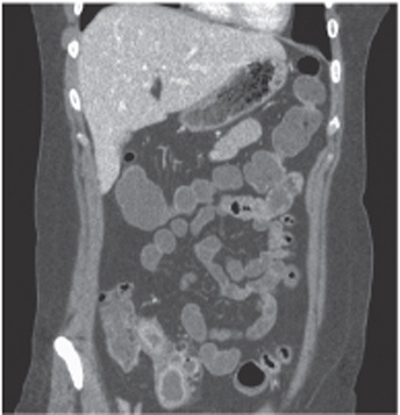
CASE 87 A 45-year-old man with chronic abdominal symptoms presents with abdominal pain, diarrhea, and weight loss. Fig. 87.1 An axial CT image shows mild enhancement and thickening of terminal ileal (arrow) and cecum walls at the ileocecal junction with mild peri-inflammatory fat stranding. Fig. 87.2 An axial CT image shows moderate, irregular wall thickening of the terminal ileum (arrow). The walls of inflamed bowel loops have a stratified appearance and present mild enhancement after intravenous administration of contrast medium. There is prominence of mesenteric vessels surrounding the inflamed bowel. Axial and coronal computed tomography (CT) images (Figs. 87.1, 87.2, and 87.3) of the abdomen obtained after oral administration of negative contrast material show diffuse, irregular wall thickening of distal small bowel loops mostly affecting the terminal ileum. There is moderate prominence of the fat surrounding the diseased bowel loops. The ileocecal junction appears affected as well, with mild cecal wall thickening. Crohn disease (regional enteritis/terminal ileitis/granulomatous ileocolitis) Crohn disease is a chronic granulomatous inflammatory condition of unknown etiology, occurring in multiple discontiguous sites of the gastrointestinal tract. Lesions may occur anywhere from the mouth to the anus but are mostly encountered in the small bowel, especially the terminal ileum (in 80%), colon (in 70%), and, less commonly, duodenum (in 20%). The small and large bowels are frequently both involved. Incidence is higher among Caucasians in their 2nd to 3rd decade, and women are slightly more affected than men. The most typical pathologic feature is the involvement of all layers of the diseased bowel.
Clinical Presentation
Radiologic Findings
Diagnosis
Differential Diagnosis
Discussion
Background
Related posts:


Stay updated, free articles. Join our Telegram channel

Full access? Get Clinical Tree


