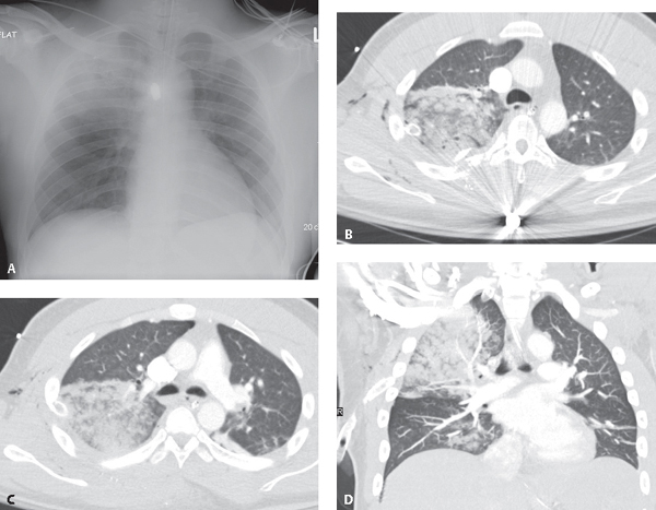
CASE 88 Young male victim of a gunshot wound to the right chest AP chest X-ray (Fig. 88.1A) demonstrates extensive right upper lobe air space disease. Metallic shrapnel fragments follow a path from the mid-clavicle to the first intercostal space and continue over the fractured third to fifth posterior ribs to a retained bullet over T4-T5. Foci of subcutaneous air overlie the right coracoid process. Axial (Figs. 88.1B, 88.1C) and coronal (Fig. 88.1D) chest CT (lung window) through the right upper lobe show the ground glass with acinar opacities and consolidation to better advantage. Note the comminuted rib fractures and subcutaneous air. A chest tube has been placed. Pulmonary Contusion Fig. 88.1 None Pulmonary contusion is equivalent to a bruise of the lung that leads to edema and blood accumulation in alveolar spaces and loss of normal lung function. Pulmonary contusions are the most common pulmonary injury after blunt thoracic trauma and are present in 17–70% of patients with severe chest trauma.
 Clinical Presentation
 Radiologic Findings
 Diagnosis
 Differential Diagnosis
 Discussion
Background
Etiology
Related posts:


Stay updated, free articles. Join our Telegram channel

Full access? Get Clinical Tree


