Case 89
 Clinical Presentation
A 38-year-old man with left lower quadrant pain. Contrast-enhanced computed tomography was performed to determine the cause.
 Imaging Findings
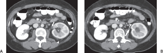
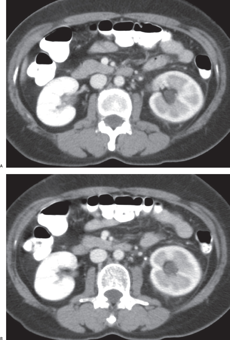
(A) Contrast-enhanced computed tomography (CT) image at the level of the kidneys shows that the left kidney (short arrow) is in the corticomedullary phase while the right kidney is in the nephrographic phase. Therefore, the passage of contrast through the left kidney is lagging passage through the right. There is mild dilatation of the collecting system on the left (asterisk). Note the mildly dilated upper ureter (long arrow). Mild perinephric stranding (arrowheads) is also seen. (B) Contrast-enhanced CT image at a level immediately inferior to that in Figure A shows the dilated ureter (arrow) ending in a hyperattenuating focus (arrowhead) whose attenuation is higher than that of any of the vessels opacified with intravenous contrast.
 Differential Diagnosis
• Obstructing left ureteric calculus:
Stay updated, free articles. Join our Telegram channel

Full access? Get Clinical Tree


