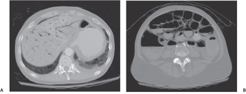
CASE 89 A 72-year-old man presents with diffuse abdominal pain. Fig. 89.1 (A) Axial CT image shows gas in the portal vein branches (peripherally located, as opposed to central gas in the pneumobilia) and free gas under the anterior abdominal wall. (B) Lower axial CT image in the same patient shows curvilinear gas collections in the wall of the distended bowel loops. Axial computed tomography (CT) images in a patient with massive bowel ischemia show the presence of hypodense curvilinear gas lucencies in the bowel wall and portal vein branches (Fig. 89.1). Bowel pneumatosis with portal venous gas The presence of gas within the bowel wall is termed pneumatosis intestinalis. Recognition of pneumatosis intestinalis in symptomatic patients is important because it may be the only finding of an underlying pathology, which may not be always obvious on imaging. Pneumatosis intestinalis can be primary (idiopathic) or secondary (due to underlying causes).
Clinical Presentation
Radiologic Findings
Diagnosis
Differential Diagnosis
Discussion
Background
Clinical Findings
Related posts:


Stay updated, free articles. Join our Telegram channel

Full access? Get Clinical Tree


