
BI-RADS Categorization | ||
Clinical Findings | right 1 | left 1 |
Ultrasound | right 1 | left 1 |
Mammography | right 3 | left 1 |
BI-RADS Total | right 3 | left 1 |
This is the imaging study of an asymptomatic woman.
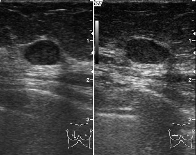
Ultrasound
Ultrasound showed a well-defined lesion measuring 9 mm in the center of the right breast, with increased distal acoustic signal. US BI-RADS right 3/left 1.
Mammography
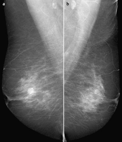
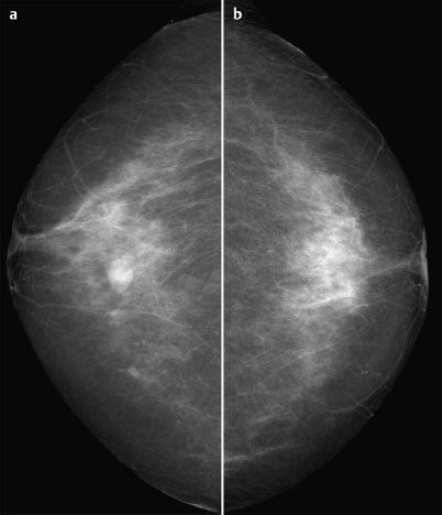
Fibroglandular parenchyma, ACR type 2. Compared to the mammograms from 2.5 years previously, a well-defined, oval, isodense lesion had increased from 0.4 cm to 1 cm in diameter. No architectural distortion. BI-RADS right 3/left 1. PGMI: CC view P; MLO view G (inframammary fold incorrectly positioned).
Procedure
A lesion increasing in size is grounds for further investigation in a 68-year-old patient, even when morphological appearance is benign. For this reason, US-guided core biopsy was performed here.
Histology
The initial diagnosis was tubular adenoma. However, after immunohistochemical work-up a diagnosis of intraductal papilloma was reached.
Further procedure
Because of the risk of malignant transformation associated with intraductal papillomas, resection of the benign lesion was recommended.
Final Histopathology
Intraductal papilloma.


Stay updated, free articles. Join our Telegram channel

Full access? Get Clinical Tree


