Case 91
Indication: Screening.
History: Unremarkable.
Risk profile: No increased risk.
Age: 53 years.

Fig. 91.1 Inspection.
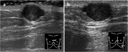
Fig. 91.2a,b Sonography.
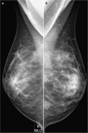
Fig. 91.3a,b Digital mammography, MLO view.
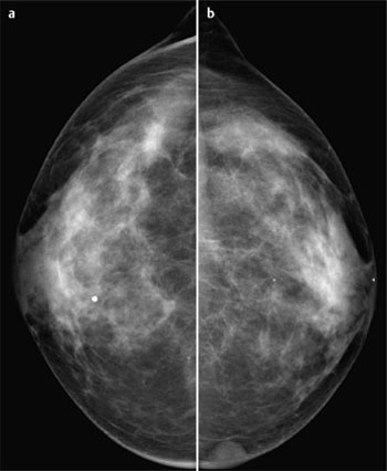
Fig. 91.4a,b Digital mammography, CC view.
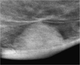
Fig. 91.5 Magnification view of the inner region of the left breast.

|
Please characterize ultrasound, mammography,
and clinical findings.
What is your preliminary diagnosis?
What are your next steps?
|


