CASE 92 A 70-year-old patient presents with constipation and hematochezia. Fig. 92.1 Axial contrast-enhanced CT image of the sigmoid colon shows an enhancing “apple core” lesion causing short segment narrowing (arrow). Fig. 92.2 Axial CT image of the upper abdomen in the same patient shows a peripherally enhancing hypodense hepatic metastasis in the liver (arrow). Also seen are perihepatic malignant ascites. Axial contrast-enhanced computed tomography (CT) images through the pelvis show an enhancing “apple core” lesion causing short segment narrowing in the sigmoid colon (Figs. 92.1, 92.2). Figure 92.2 shows a hypodense peripherally enhancing hepatic metastasis in the same patient, along with malignant perihepatic ascites. Adenocarcinoma sigmoid colon with hepatic metastasis Colorectal cancers are the second most common cause of cancer-related deaths in the United States and Europe. The sigmoid colon is the second most common site of colonic malignancy following the rectum.
Clinical Presentation
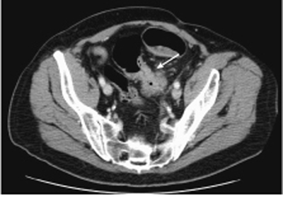
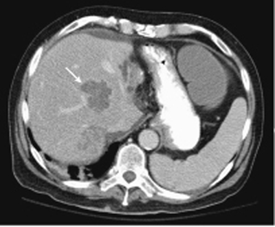
Radiologic Findings
Diagnosis
Differential Diagnosis
Discussion
Background
Clinical Findings
Related posts:


Stay updated, free articles. Join our Telegram channel

Full access? Get Clinical Tree


