Case 93
Indication: Newly detected microcalcifications in the left breast.
History: Unremarkable.
Risk profile: Breast cancer in mother at the age of 60 years.
Age: 56 years.
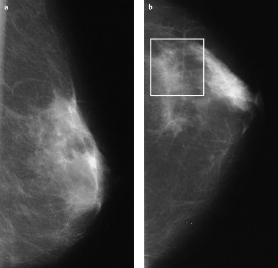
Fig. 93.1a,b Conventional mammography of the left breast, MLO and CC views [imaging not performed by authors].
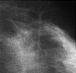
Fig. 93.2 Magnification view of the outer quadrants of the left breast (CC).
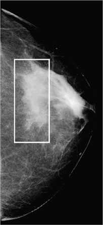
Fig. 93.3 Digital mammography of the left breast, CC view, in preparation for intervention 5 weeks later.
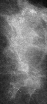
Fig. 93.4 Magnification view (CC) of the left breast (see section marked in 93.3).
Clinical Findings
No findings.
Ultrasound (not shown)
Unremarkable.

|
Please characterize the mammography from 5 weeks earlier and the current images. What is your preliminary diagnosis? What are your next steps? |


