Case 93
 Clinical Presentation
A 48-year-old woman with pelvic pressure on the left.
 Imaging Findings
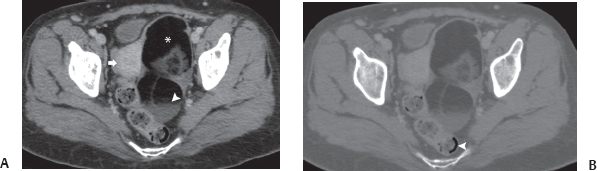
(A) Contrast-enhanced computed tomography of the pelvis obtained at the level of the uterus shows the body of the uterus (arrow) to be pushed to the right by a large mass (asterisk) on the left side of the pelvis. The mass has very hypoattenuating contents mixed with areas of soft-tissue attenuation. A fluid level is seen in the dependent portion of the mass (arrowhead). (B) When the same image is viewed in wide window, the contents of the mass become grayer and the gas in the bowel (arrowhead) remains black, thus showing that the hypoattenuating areas in the mass consist of fat.
 Differential Diagnosis
• Ovarian dermoid cyst: A large mass in a woman’s pelvis that contains fat and soft tissue is a dermoid cyst unless proven otherwise. The fat is characteristically significantly more hypoattenuating than adipose tissue, and fat–fluid levels are characteristic.
• Pelvic liposarcoma:
Stay updated, free articles. Join our Telegram channel

Full access? Get Clinical Tree


