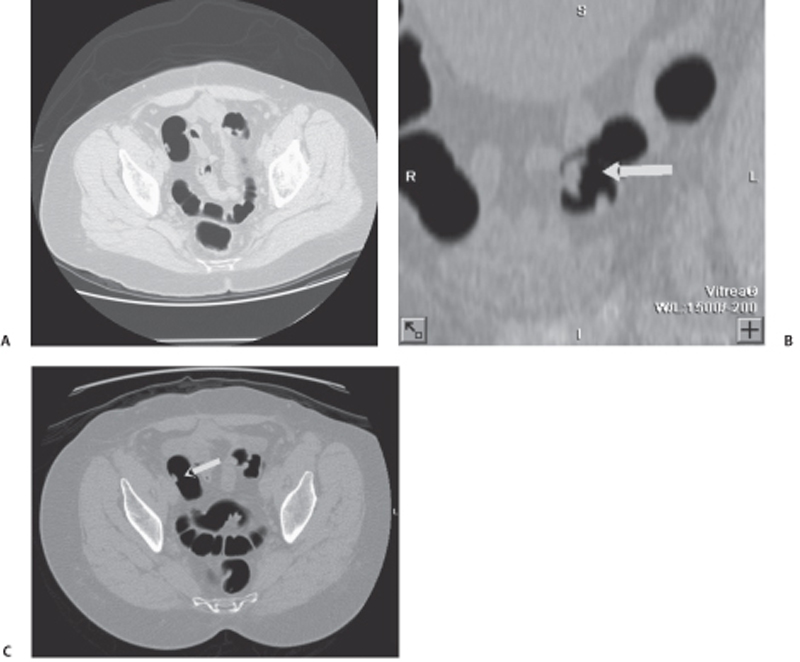
CASE 93 A 69-year-old man presents with abdominal pain and gastrointestinal (GI) bleeding. Fig. 93.1 (A–C) CT colonoscopy with insufflated carbon dioxide in the colon. Axial CT images of the pelvis show a small isodense lesion protruding in the wall of the sigmoid colon, consistent with a sessile polyp (arrow). Axial images from computed tomography (CT) colonography show a small isodense protruding lesion arising from the wall of the sigmoid colon (Fig. 93.1). Sigmoid colonic polyp
Clinical Presentation
Radiologic Findings
Diagnosis
Differential Diagnosis
Discussion
Background
Related posts:


Stay updated, free articles. Join our Telegram channel

Full access? Get Clinical Tree


