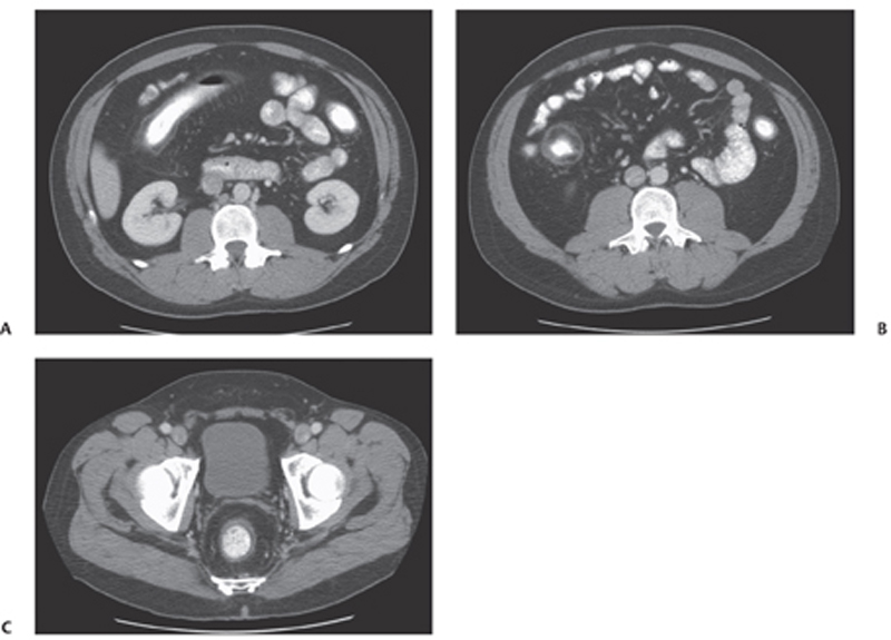
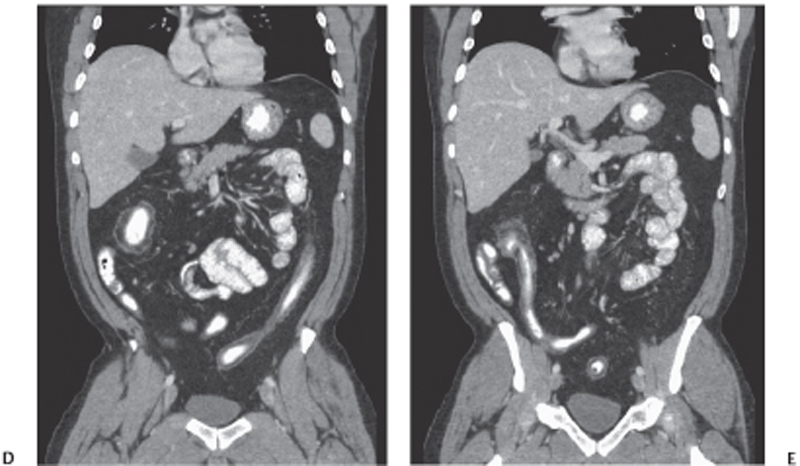
CASE 94 A 45-year-old man presents with abdominal cramps and rectal bleeding. Fig. 94.1 (A–C) Axial CT images of the abdomen show mild diffuse wall thickening of the ascending colon with surrounding peri-inflammatory changes. Multiple small lymph nodes are noticed. The thickened wall presents a typical stratified appearance with negative attenuation (fatty halo sign) of the middle layer and mild hyperdensity of the inner and outer layer. No fluid collections are seen. (D,E) Coronal CT images of the abdomen show the typical foreshortened, narrowed, and ahaustral appearance of the affected colon in ulcerative colitis. All the segments of the large bowel are similarly involved. Note the involvement of the terminal ileum (backwash ileitis). Axial and coronal computed tomography (CT) images of the abdomen obtained after administration of positive oral contrast medium (Fig. 94.1) show diffuse wall thickening of the entire large bowel. The mesentery presents mild fat stranding with multiple small lymphadenopathies mostly located in the right quadrant close to the ascending colon. There are no fluid collections. Ulcerative colitis
Clinical Presentation
Radiologic Findings
Diagnosis
Differential Diagnosis
Discussion
Background
Related posts:


Stay updated, free articles. Join our Telegram channel

Full access? Get Clinical Tree


