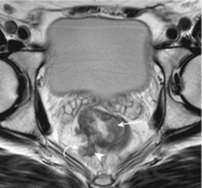
CASE 95 A 64-year-old man complains of bloody diarrhea. Fig. 95.1 Axial T2-weighted MRI image of the pelvis shows ill-defined circumferential thickening of the rectum due to a low signal intensity tumor (arrow). There is extension of the tumor into the perirectal fat (arrowhead), making it a T3 disease, as well as an adjacent metastatic lymph node (curved arrow). Axial T2-weighted magnetic resonance (MR) image of the pelvis shows ill-defined circumferential thickening of the rectum. There is soft tissue extension into the perirectal fat as well as an adjacent lymph node (Fig. 95.1). Rectal adenocarcinoma with perirectal extension of tumor Colorectal carcinoma is the second most common cancer in the United States. Approximately one third of these cancers occur in the rectum. Ninety-eight percent of rectal cancers are adenocarcinomas, and they usually arise from adenomatous polyps.
Clinical Presentation
Radiologic Findings
Diagnosis
Differential Diagnosis
Discussion
Background
Related posts:


Stay updated, free articles. Join our Telegram channel

Full access? Get Clinical Tree


