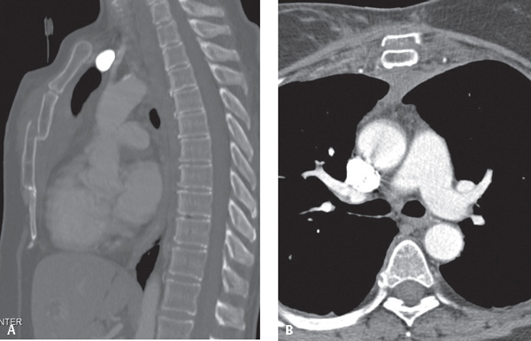
CASE 96 59-year-old unrestrained driver involved in a high-speed MVC with complaints of chest pain. The vehicle’s steering wheel was severely deformed in the collision. Contrast-enhanced sagittal MIP (bone window) (Fig. 96.1A) and axial (mediastinal window) (Fig. 96.1B) chest CT shows a markedly depressed mid-sternal body fracture; associated peri-sternal, para-sternal, and retrosternal hematoma; and soft-tissue stranding overlying the sternum. Note the preserved fat plane between the retrosternal hematoma and the aorta. Sternal Fracture None Fig. 96.1 The incidence of sternal fracture as a result of motor vehicle collisions ranges between 3% and 10%, although the incidence may decrease with airbags. The most common location for sternal fractures is within 2 cm of the sternal-manubrial joint. Most fractures are associated with a retrosternal hematoma (Fig. 96.1B). Simple sternal fractures are usually benign. Depressed, segmental sternal fractures have an increased association with myocardial contusions, traumatic hemopericardium, coronary vessel lacerations, thoracic aorta lacerations, tracheobronchial tears, thoracic spine fractures (see Case 99), and head trauma.
 Clinical Presentation
 Radiologic Findings
 Diagnosis
 Differential Diagnosis
 Discussion
Background
Etiology
Related posts:


Stay updated, free articles. Join our Telegram channel

Full access? Get Clinical Tree


