CASE 97 A 45-year-old Armenian man presents with 2 days of severe left upper abdominal pain and fever. He has had similar episodes every few months since childhood. Sometimes these episodes are associated with nausea and vomiting, but there is no associated diarrhea. The episodes usually last a few days and are self-limited. His past medical history is otherwise significant for remote appendectomy and small bowel obstruction with exploratory laparotomy and lysis of adhesions. Fig. 97.1 (A) Axial and coronal CT at the time of initial presentation shows pericolonic and perienteric soft tissue inflammatory stranding in the left upper abdomen. (B) CT performed 2 months later shows mesenteric vascular engorgement, omental and mesenteric edema, and fluid in the left paracolic gutter. (C) A third CT scan performed 2 months after the second scan shows right nephromegaly and new asymmetric right perirenal fat stranding; a 4 mm distal right ureteral stone was diagnosed (not shown). Notice the absence of inflammatory changes in the peritoneal cavity. Axial and coronal computed tomography (CT) scans at the time of initial presentation (Fig. 97.1A) show pericolonic and perienteric soft tissue inflammatory stranding in the left upper abdomen. CT performed 2 months later (Fig. 97.1B) shows mesenteric vascular engorgement, omental and mesenteric edema, and fluid in the left paracolic gutter. A third CT scan performed 2 months after the second (Fig. 97.1C
Clinical Presentation
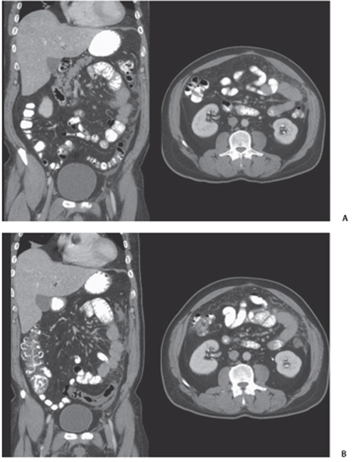
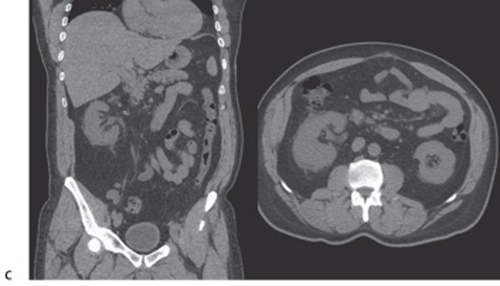
Radiologic Findings
![]()
Stay updated, free articles. Join our Telegram channel


Full access? Get Clinical Tree


