
Clinical Findings | right 1 | left 1 |
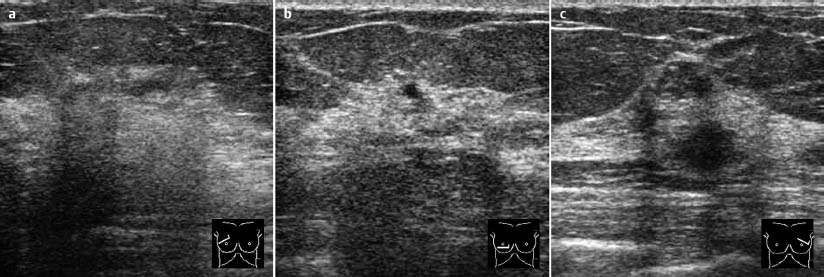
Ultrasound | right 2 | left 2 |
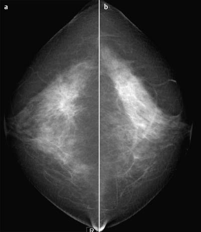
Mammography | right 4 | left 1 |
BI-RADS Total | right 4 | left 2 |
The images presented were obtained in the context of a screening examination.
Ultrasound
Bilaterally ultrasound showed glandular tissue with macrocysts. No signs of malignancy in the upper outer quadrant of the right breast or elsewhere. US BI-RADS right 2/left 2.
Mammography
Extremely dense, asymmetric glandular texture (ACR type 4) with signs of tissue retraction in the upper outer quadrant of the right breast (architectural distortion, Fig. 98.4). Margins of a tumor could not be distinctly established. No microcalcifications. No further unusual findings. BI-RADS right 4/left 1. PGMI: CC view P; MLO view G (inframammary fold).
Procedure
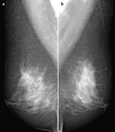
Histological investigation (here by MR-guided vacuum core biopsy). Diagnostic MR mammography (Fig. 98.5).
Histology of the core biopsy (right breast)
Invasive ductal carcinoma G2 developed from a base of DCIS (high grade, without necrosis).
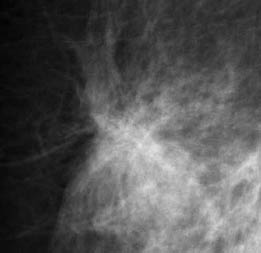
Fig. 98.4 Magnification view of the cranial region of the right breast.
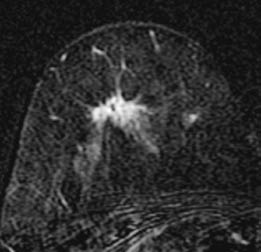
Fig. 98.5 Contrast-enhanced MR mammography of the right breast.
Histology
Invasive ductal carcinoma measuring 14 mm with an extensive intraductal component.
IDCpT1c+EIC, pN0(0/21),G2.

Stay updated, free articles. Join our Telegram channel

Full access? Get Clinical Tree


