Case 98
 Clinical Presentation
A 44-year-old woman with a history of recurrent episodes of left flank pain. She underwent a hysterectomy a few years previously.
 Imaging Findings
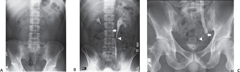
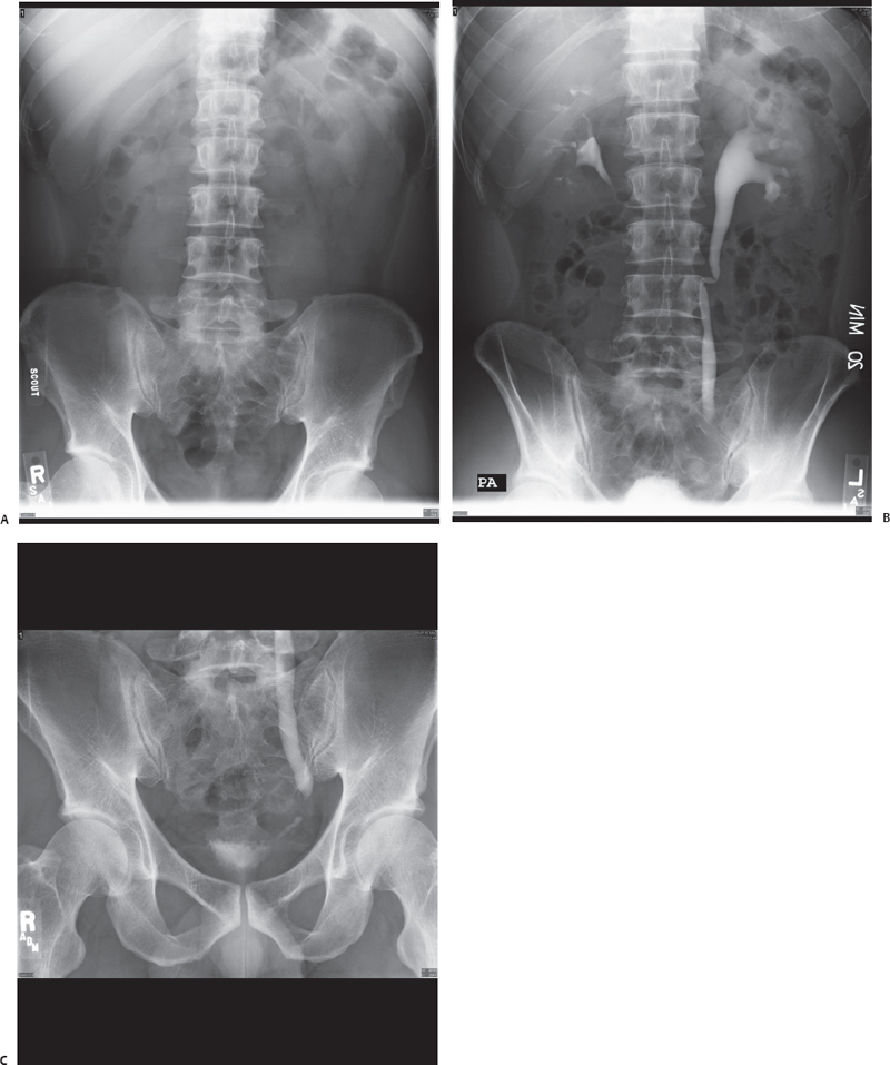
(A) Scout view of the abdomen shows no urinary tract calcifications. However, the scout view is incomplete because the symphysis pubis has not been included. Actually, this was one of two scout images. The other included the whole pelvis and was also normal. (B) Twenty-minute intravenous pyelogram (IVP) image shows left hydronephrosis (asterisk) and hydroureter (arrow). The ureter is redundant (arrowhead), suggesting that this is a long-standing abnormality. The transitional zone has not been reached. Therefore, the level and cause of the obstruction cannot be commented on. (C) Postvoid IVP image shows smooth tapering of the dilated left ureter into a smooth, narrowed segment (arrow). The terminal portion (arrowhead) of the ureter is normal. There is no significant postvoid residual urine in the urinary bladder (asterisk). The outline and mucosal pattern of the urinary bladder are normal.
Stay updated, free articles. Join our Telegram channel

Full access? Get Clinical Tree


