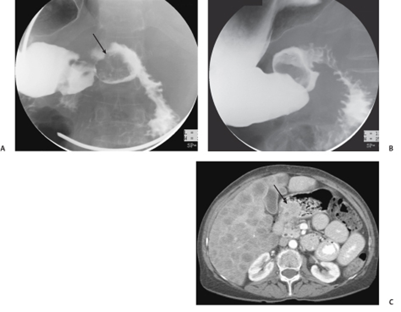
CASE 98 A 59-year-old woman with a history of breast cancer presents with a complaint of abdominal pain and weight loss. Fig. 98.1 (A,B) Upper gastrointestinal (GI) study shows a lobulated filling defect in the duodenal bulb (arrow). There is no wall irregularity. (C) CT scan performed in the same patient shows multiple rim-enhancing lesions in the liver, likely representing metastasis with central areas of necrosis. There is an enhancing mass in the antrum of the stomach (arrow). Given the appearance on the upper GI series and the CT appearance, the primary lesion is likely a pedunculated lesion arising from the antrum and herniating into the duodenal bulb. Upper gastrointestinal (GI) study shows a lobulated filling defect in the duodenal bulb. Computed tomography (CT) scan performed in the same patient shows multiple rim-enhancing lesions in the liver, likely representing metastasis with central areas of necrosis. There is an enhancing mass in the antrum of the stomach (Fig. 98.1). Pedunculated gastric carcinoid, type III (sporadic subtype)
Clinical Presentation
Radiologic Findings
Diagnosis
Differential Diagnosis
Discussion
Background
Related posts:


Stay updated, free articles. Join our Telegram channel

Full access? Get Clinical Tree


