Acknowledgment
The authors would like to thank Shundra Dinkins, Toni Acfalle, and Dana Murphy for their expertise in preparation of this manuscript.
The authors also would like to acknowledge Jozef M. Brozyna, Suma Chandra, Hank K. Chen, Gina D. Tran, and Dimitri A. Gagarin for their contributions to this chapter, which were published in previous editions of this book.
Historical Highlights of Endovascular Therapy
- •
British dentist Charles Stent develops a plastic material for taking mouth impressions (i.e., creates a “scaffold”) (1856).
- •
Wilhelm Conrad Roentgen’s discovery of x-rays on November 8, 1895, makes image guidance possible ( Figs. 1.1 and 1.3 ).

Fig. 1.1
Photograph of Roentgen taken in 1906 while he was director of the Institute of Physics at the University of Munich.
From Eisenberg RL. Radiology: An Illustrated History . St Louis: Mosby–Year Book; 1992:38.

Fig. 1.2
First roentgen photograph of Mrs. Roentgen’s hand.
From Glasser O. Wilhelm Conrad Röntgen and the Early History of the Roentgen Rays . Springfield, IL: Charles C Thomas; 1933.

Fig. 1.3
Roentgen’s first communication. Left, First page of handwritten manuscript (1895). Middle, First page of published article on new type of ray. Right, Front cover of a reprint of initial paper.
From Eisenberg RL. Radiology: An Illustrated History . St Louis: Mosby–Year Book; 1992:25.
- •
Sven Ivar Seldinger develops percutaneous vascular catheterization (1952) ( Figs. 1.4 and 1.5 ).

Fig. 1.4
Sven Ivar Seldinger.
From Eisenberg RL. Radiology: An Illustrated History . St Louis: Mosby–Year Book; 1992: 442.

Fig. 1.5
Seldinger technique (1953). (A) Equipment. Stiletto is removed and leader inserted through needle and catheter. (B) Diagram of technique used: (1) artery punctured and needle pushed upward, (2) leader inserted, (3) needle withdrawn and artery compressed, (4) catheter threaded onto leader, (5) catheter inserted into artery, (6) leader withdrawn.
From Seldinger SI. Catheter replacement of the needle in the percutaneous arteriography: a new technique. Acta Radiol. 1953;39:368–76.
- •
Charles Dotter accomplishes percutaneous revascularization with coaxial dilators (1964) ( Figs. 1.6 and 1.7 ).

Fig. 1.6
Dotter coaxial Teflon catheter system consisting of 12F catheter with tapered and beveled tip over inner 8F catheter with 0.044-inch guidewire.
From Waltman AC, Greenfield AJ, Athanasoulis CA. Transluminal angioplasty: general rules and basic considerations. In: Athanasoulis CA, Greene RE, Pfister RC, Roberson GH, eds. Interventional Radiology . Philadelphia: WB Saunders; 1982.

Fig. 1.7
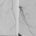
First percutaneous transluminal angioplasty (1964). (A) Control arteriogram showing segmental narrowing, with threadlike lumen of left superficial femoral artery in region of adductor hiatus. (B) Study immediately after dilation with catheter having an outer diameter of 3.2 mm. (C) Three weeks after transluminal dilation, lumen remains open. Clinical and plethysmographic studies indicated continuing patency more than 6 months later.
From Dotter CT, Judkins MP. Transluminal treatment of arteriosclerotic obstruction. Circulation 1964;30:654–70.
- •
Charles Dotter performs transcatheter embolotherapy to control acute upper gastrointestinal bleeding (1970).
- •
Lazar Greenfield pioneers caval interruption with a vena cava filter (1973) ( Fig. 1.8 ).

Fig. 1.8
Kimray-Greenfield filter.
From Dedrick CG, Novelline RA. Transvenous interruption of the inferior vena cava. In: Athanasoulis CA, Greene RE, Pfister RC, Roberson GH, eds. Interventional Radiology . Philadelphia: WB Saunders; 1982.
- •
Andreas Gruentzig performs percutaneous angioplasty (1977) ( Figs. 1.9 and 1.10 ).

Fig. 1.9
Percutaneous transluminal coronary angioplasty (1979). (A) Stenosis of coronary artery. (B) Double-lumen balloon catheter is introduced with guiding catheter positioned at orifice of left or right coronary artery. At tip of dilating catheter is a short soft wire to guide catheter through vessel. Proximal to wire is a side hole connected to main lumen of dilating catheter. This lumen is used for pressure recording and injection of contrast material. Dilating catheter is advanced through coronary artery with balloon deflated. (C) Balloon is inflated across stenosis to its predetermined maximal outer diameter, thereby enlarging lumen. After balloon deflation, catheter is withdrawn.
From Grüntzig AR, Senning A, Siegenthaler WE. Non-operative dilation of coronary artery stenosis. Percutaneous transluminal coronary angioplasty. N Engl J Med. 1979;301:61–8. Copyright 1979 Massachusetts Medical Society. All rights reserved.

Fig. 1.10
Transluminal dilation of coronary artery stenosis (1978). Left, Initial angiogram in 43-year-old man with severe angina pectoris reveals severe stenosis of main left coronary artery. Middle, After passage of dilation catheter, distensible balloon segment was inflated twice to a maximum outer diameter of 3.7 mm. Right, Postprocedure angiogram showing good result without complications.
From Grüntzig AR. Transluminal dilation of coronary artery stenosis. Lancet. 1978;1:263.
- •
Tetsuro Kato performs chemoembolization. (1981).
- •
Julio Palmaz develops the endovascular balloon-expandable stent (1985).
- •
Juan C. Parodi, Julio C. Palmaz, and H.D. Barone develop stent-grafts (1990).
Endovascular Milestones
November 8, 1895, was a day of historical significance in endovascular medicine. In a physics laboratory in southern Germany, Wilhelm Conrad Roentgen accidentally discovered x-rays. The mysterious rays illuminated medical science, and radiology was born. From this serendipitous discovery, image-guided minimally invasive procedures evolved to provide patients with therapeutic options in the management of vascular and nonvascular diseases.
This chapter is neither comprehensive nor complete. Of the many potential technologic advances, arterial and venous endovascular therapy is the focus in this section. Later in the chapter, the history of nonvascular interventions is covered.
Arterial Endovascular Therapy: Revascularization and Vessel Reconstruction
An English dentist, Dr. Charles Stent, developed a thermoplastic material for taking impressions of toothless mouths in 1856. Thus the “stent” may be considered a “scaffold.” For purposes of this discussion, a stent is used for reconstruction in the vascular (or nonvascular) systems.
Percutaneous vascular catheterization as a viable endovascular technique was described in June 1952, when Sven Ivar Seldinger presented his idea of replacing an arteriography needle with a catheter. Translumbar aortography and direct carotid puncture were effectively relegated to historical descriptions in textbooks.
The technique of percutaneous revascularization advanced rapidly in 1964, when Charles Dotter used coaxial “pencil-point” dilators to treat a superficial femoral artery stenosis. The era of image-guided revascularization of the lower extremities had begun.
Although Dotter was successful in dilating femoral arterial stenoses, the use of coaxial pencil-point dilators (van Andel Catheters [Cook Medical, Bloomington, IN]) required progressive enlargement of the percutaneous puncture site. Development of the angioplasty balloon by Andreas Gruentzig in 1977 resulted in another major step forward that allowed the percutaneous arterial entry site to be kept to a minimum ( Figs. 1.9 and 1.10 ). Early balloon designs were hampered by uneven balloon dilation and frequent rupture. Such events often led to pseudoaneurysm formation or dissections and resulted in vessel thrombosis.
The arterial metallic stent was invented in the 1980s by Julio Palmaz at the University of Texas Health Science Center in San Antonio. Dr. Palmaz described his stent in 1985 and continued work on his device in 1986. Later developments included the use of a stent design consisting of a single stainless steel tube with parallel staggered slots in the wall. When the stainless steel tube was expanded, the slots formed diamond-shaped spaces that resisted arterial compression. This design became the first stent approved by the US Food and Drug Administration (FDA) for vascular use.
Stanley Baum and Moreye Nusbaum pioneered catheter embolization in the mid-1960s for treating acute gastrointestinal bleeding. In 1970 Charles Dotter reported utilizing an autologous clot as the embolic agent to control acute upper gastrointestinal bleeding by selective embolization of the right gastroepiploic artery in a patient who was a poor surgical candidate. Robert White used this technique in 1974 to control bleeding duodenal ulcers when the hemorrhage was unresponsive to intra-arterial injections of vasopressin.
Chemoembolization was largely pioneered by the Japanese urologist Kato in 1981. Kato used particles about 200 μm in size to demonstrate that chemoembolization with microcapsules containing chemotherapeutic agents was superior to local intraarterial injection of antitumor agents.
Stent-grafts, pioneered by Juan Parodi and Charles Dotter, became the major impetus for future endovascular reconstruction procedures used to exclude an aneurysm, close an arteriovenous fistula, and reconstruct the central lumen of a dissected vessel. Specifics of the history of endovascular grafts are worth mentioning. The technology has fundamentally changed the management of diseases of the abdominal and thoracic aorta.
The first abdominal aortic aneurysm (AAA) was described by Andreas Vesalius in the 16th century. By the 1800s, aortic ligation had become the surgical procedure of choice when attempting to treat iliac and abdominal aortic aneurysms. The longest survivor was a patient of Keen in 1899, who survived 48 days after aortic ligation at the diaphragm for a ruptured AAA. This surgery was deemed a great success because previous aortic ligations for iliac aneurysms performed by Astley Cooper in 1817 and J.H. James in 1829 resulted in patient death within 48 hours. As the years passed, many others attempted aortic ligations, and most met similar results; the most common mechanism of failure was ligature erosion into the aorta and massive hemorrhage. In April 1923, more than 100 years after Cooper’s first attempts, Rudolph Matas performed the first successful aortic ligation on a patient with a syphilitic aorta and bilateral common iliac aneurysms, using cotton tape. The patient survived for 17 months before succumbing to tuberculosis.
The subsequent evolution of aortic aneurysm repair involved many varied techniques. Physicians experimented with different variations of aortic ligation, wires to induce thrombosis, and, most notably, reactive polyethylene cellophane wrapping of aneurysms. This particular technique was performed on Albert Einstein in 1949; however, he eventually died of an AAA rupture 6 years later. In 1951 the first homograft was used for AAA repair by Charles DuBost, only to be superseded by nylon and then polytetrafluoroethylene grafts shortly thereafter. Javid and Creech first introduced surgical endoaneurysmorrhaphy in 1961, greatly reducing the high mortality rates associated with aneurysm excision and graft repair.
With the advent of minimally invasive techniques for the treatment of AAAs, the evolution of aneurysm repair changed dramatically. In the late 1970s Juan Parodi had begun thinking about endovascular aneurysm repair (EVAR) during his vascular surgery fellowship at the Cleveland Clinic. After creating many rudimentary prototypes of varying designs, he became convinced that AAA repair was possible without laparotomy. However, he continuously ran into two problems: (1) the mechanics of delivering a minimally invasive system into the precise location needed, and (2) the absence of a fastening device that would take the place of surgical sutures in securing the graft in place and creating a seal between the intravascular graft and vessel wall.
The solution to these challenges came when Parodi met Julio Palmaz at the Transcatheter Cardiovascular Therapeutics meeting in Washington, DC, in 1988. Palmaz was attending to discuss a new stent design. Parodi was informed that Palmaz was presenting and thus was encouraged to attend. It became apparent to Parodi that the Palmaz design was precisely what he was looking for: a device with a high radial force (i.e., a balloon-expandable stent) that was deployed using endovascular techniques and would provide placement, precision, a secure anchor, and an aortic wall seal that his EVAR concept required. Palmaz was initially apprehensive about joining the project because of his many professional commitments and the perceived limitations of his device in the aorta. However, he later admitted that he envisioned his stent playing a role in aneurysm repair as early as 1986. Ultimately, he decided to join efforts with Parodi. Work then began on the first EVAR in both Buenos Aires, where Parodi worked at the Instituto Cardiovascular de Buenos Aires (ICBA), and San Antonio, where Palmaz was Chief of Special Procedures at the University of Texas Health Science Center. During their efforts to perfect the device and the technique, Parodi was searching for an ideal candidate for the first EVAR.
On September 7, 1990, two patients were to undergo the new procedure, to be performed by both Parodi and Palmaz at the cardiac catheterization laboratory of ICBA. During the first procedure, the proximal portion of the stent was deployed just inferior to the renal arteries, and the distal portion placed just superior to the aortic bifurcation. The postprocedure angiogram displayed a completely excluded aneurysm, concluding the first EVAR procedure. The second patient was converted to traditional surgical AAA repair, because the proximal portion of the stent was deployed too low in the aorta, and the distal end of the graft ended in one common iliac artery. It was not until later that Parodi realized that the reflection of pressure waves from the aortic bifurcation and iliac arteries would cause an endoleak and failure of a single stent device.
This initial experience provided Parodi and Palmaz with clinical insight into patient benefits, such as improvements in recovery time and quality of life, using an endovascular approach. The first patient quickly recovered and was discharged without complications, whereas the second remained intubated in the intensive care unit. Additionally, the second case provided the team with a possible mechanism of failure for their new procedure and delivery system. Over time, the procedure, delivery system, and methods of Parodi’s team evolved. By December 1992, 24 patients had undergone EVAR by one of three techniques that had evolved since the first case, the most common being an aorto-uni-iliac approach with contralateral common iliac embolization and femoral-femoral bypass. Before this, an interventional radiologist from Buenos Aires, Claudio Schonholz, had also joined the international team and played a significant role in the first EVAR performed in the United States.
In August 1992 the vascular surgery department at Montefiore Medical Center in Bronx, New York, was consulted on a 76-year-old male patient with a 7.5-cm infrarenal AAA with several comorbidities that suggested he was not a candidate for open surgical repair. Frank Veith and Michael Marin quickly contacted Parodi and began discussing the logistics involved in traveling to South America, learning the procedure, and treating the aneurysm endovascularly. After several meetings, and convincing executives at Johnson & Johnson (which was producing the Palmaz stent) to allow them to use a large Palmaz stent for the procedure (no FDA Investigational Device Exemption existed for it yet), they were ready to proceed. On November 23, 1992, Parodi, Schonholz, Veith, Marin, and Jacob Cynamon performed the first EVAR in the United States, using a 22-mm Dacron prosthesis sewn over a large Palmaz-like stent. The patient was discharged several days later and remained symptom free until his death 9 months later. Death was due to problems unrelated to the EVAR procedure.
Although Parodi is often credited with the first EVAR, it is important to mention that a Ukrainian surgeon, Nicholas Volodos, published an article in 1986 describing the endovascular repair of a traumatic thoracic aortic aneurysm with a homemade stent-graft. Also of note, Harrison Lazarus was awarded a US patent in 1988 for an endovascular stent-graft that eventually served as the basis for the Guidant (Indianapolis, IN) Ancure device.
With advancements in technology and refinement of techniques, EVAR has become for selected patients the standard of care for treatment of AAAs. By 2013, approximately 80 percent of Medicare patients with AAA in the United States were repaired endovascularly. There are an estimated 1.1 million Americans between the ages of 50 and 84 years diagnosed with an AAA, and more than 15,000 deaths annually due to abdominal aortic aneurysms, so it is no surprise that this minimally invasive treatment option continues to evolve.
Venous Endovascular Therapy: Caval Interruption
Treatment of venous thromboembolic disease has also rapidly evolved. Deep venous thrombosis is one of the major causes of morbidity and mortality worldwide. “Caval interruption” with a filter was initially performed via surgical cut-down by Mobbin and Uddin. In 1973 Lazar Greenfield introduced a cone-shaped surgically placed vena cava filter (Boston Scientific Corporation, Marlborough, MA). Greenfield pioneered his work with the Kimray Corporation. This device was first deployed percutaneously in 1984. The diameter of the sheath for the filter was 29F (outer diameter [OD]). Later developments reduced Greenfield’s percutaneous puncture size from 29F (OD) to 16F (OD).
Because all permanent vena cava filters are associated with complications, including caval thrombosis (3%–40%), recent advances have provided patients at risk for venous thromboembolic disease with optional devices, such as vena cava filters that may be permanent or removed, the latter when the patient’s “window of vulnerability” has clinically passed. Removable devices were historically called temporary filters and were classified as either tethered or retrievable . A more accurate term in current clinical use is an optional filter . Optional implies that the device may either be a permanent implant or be used for a short interval. A tethered inferior vena cava (IVC) filter consists of a filter attached to a central venous catheter. A retrievable IVC filter is a device deployed in the IVC that attaches to the IVC wall with hooks but has no external tether. (The FDA approved use of the first tethered temporary vena cava filter in the United States in the early 1990s.) This tethered device (Tempofilter [B. Braun, Evanston, IL]), placed in a young trauma patient, remained in place for 13 days and trapped a large embolus, thereby preventing a potentially life-threatening pulmonary embolic event.
The FDA approved optional vena cava filters in the United States in 2003. The Recovery Filter (Bard Peripheral Vascular, Tempe, AZ), a permanent implant with the “option to remove” at any future date, was first deployed in the United States in July 2003. Since 2003 the Günther Tulip (Cook Medical), the OPTEASE (Cordis Endovascular, Warren, NJ), and other filters have also received FDA approval as optional vena cava filters. Thus, management of venous thromboembolic disease has further evolved with newer therapeutic options for patients who are at risk for pulmonary emboli but have a contraindication to or have had a complication of anticoagulant therapy.
Summary
Arterial and venous endovascular procedures continue to progress rapidly. Future developments that might take place include continued use of mechanical devices in the vascular system, combination therapies to rapidly lyse and remove residual thrombi, further design modifications in endovascular devices to reduce the risk of metal fatigue/fracture, and the application of newer technologies to reduce intimal hyperplasia (e.g., drug-eluting stents). Such improvements will, one hopes, provide solutions for some of the limitations of our current technology.
Related posts:

Stay updated, free articles. Join our Telegram channel

Full access? Get Clinical Tree


