Overview
Location
• The aorta originates at the left ventricle of the heart then ascends posterior to the pulmonary artery; it arches to the left then descends (thoracic aorta) posterior to the diaphragm into the retroperitoneum (portion of abdominopelvic cavity posterior to the peritoneal sac) of the abdominal cavity (abdominal aorta).
• The abdominal aorta is vertically orientated in the body. It descends, anterior to the spine, just to the left of the midline then bifurcates into the common iliac arteries anterior to the body of the fourth lumbar vertebra.
Anatomy
• The largest artery in the body
• Consists of three muscle layers:
1. Intima (innermost)
2. Media (middle)
3. Adventitia (outer)
• 3 anterior branches:
1. Celiac artery (CA) trunk: also known as “celiac axis,” branches into the left gastric artery, common hepatic artery, and splenic artery
2. Superior mesenteric artery (SMA): just inferior to the CA, divides into several small arteries that supply the ascending portion of the colon and largest portion of the small intestine
3. Inferior mesenteric artery (IMA): inferior to the SMA and renal arteries, divides into several small arteries that supply the transverse and descending portions of the colon and the rectum
• 2 lateral branches:
1. Right renal artery (RRA): located a few centimeters within the origin of the SMA, it courses anterior to the spine and posterior to the inferior vena cava in route to the right kidney
2. Left renal artery (LRA): located a few centimeters within the origin of the SMA, it courses left lateral to the spine and posterior to the tail of the pancreas in route to the left kidney
• Size is normal up to 3 cm in diameter, gradually tapering toward the bifurcation
• Can be very tortuous (marked by twists, turns, or bends)
Physiology
• Supplies the organs, bones, and connective structures of the body with oxygen and nutrient-rich blood.
Sonographic Appearance
• Anechoic lumen surrounded by bright, echogenic walls.
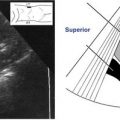
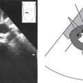
• Because the aorta is vertically orientated in the body, longitudinal and long axis views are seen in coronal scanning planes and sagittal scanning planes as demonstrated in the following image. The aorta appears like a large long tube, anechoic with bright walls, immediately anterior to the spine, and immediately posterior to the esophageal gastric junction, crus of the diaphragm, SMA, and splenic artery.

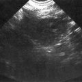
• Short axis (or axial) sections of the aorta are seen in transverse scanning planes. The following transverse scanning plane image shows how easy it is to recognize the short axis section of the aorta in the posterior portion of the image, immediately anterior and just to the left of the spine. It appears large, round, and anechoic with bright walls.

Note the axial section of the SMA branch. It appears “separate” from the aorta because following the rise of its trunk from the aorta’s anterior wall, it runs in front of, or anterior to, and parallel to the aorta. The space left between the aorta and SMA affords passage of the left renal vein (LRV) seen here in longitudinal section, on its way to the inferior vena cava (IVC).
• The proximal (closest to origin) portion of the abdominal aorta is described as the area seen inferior to the diaphragm and superior to the CA trunk; it lies anterior to the spine and posterior to the esophageal gastric junction and liver.
• The mid portion of the abdominal aorta is seen from the CA trunk running along the length of the SMA; it lies anterior to the spine and posterior to the CA, SMA, splenic artery, splenic vein, body of the pancreas, a portion of the stomach, and the liver.
• The distal (farthest from origin) portion of the abdominal aorta is described as inferior to the SMA and superior to the bifurcation; it lies anterior to the spine and posterior to the bowel.
Normal Variants
• Normal variations of the abdominal aorta are rare but include:
• Markedly tortuous
• May lie on the right side of the inferior vena cava
• May include an anterior pulmonary branch close to the CA trunk
Preparation
Patient Prep
• The patient should fast for at least 6 to 8 hours before the ultrasound study.
• If the patient has eaten, still attempt the examination.
Transducer
• 3.0 MHz or 3.5 MHz.
• 5.0 MHz for thin patients.
Breathing Technique
• Normal respiration.
• Deep, held respiration.
Patient Position
• Supine.
• Right lateral decubitus, left lateral decubitus, left posterior oblique, right posterior oblique, or sitting semierect to erect as needed.
Abdominal Aorta Survey Steps
Abdominal Aorta • Longitudinal Survey
Sagittal Plane • Transabdominal Anterior Approach
1. Begin scanning with the transducer perpendicular, at the midline of the body, just inferior to the xiphoid process of the sternum.

2. Slightly move or angle the transducer to the patient’s right and identify the longitudinal, anechoic section of the distal inferior vena cava (IVC), just posterior to the liver.

3. Return to the midline and slightly move or angle the transducer to the patient’s left and identify the longitudinal, anechoic section of the proximal aorta just posterior to the liver.

4. While viewing the proximal aorta, it may be necessary to slightly twist/rotate the transducer, first one way, then the other, to resolve the longest sections and long axis of the aorta. Additional slight twists can help reveal the proximal aorta’s anterior branches, the CA, and the SMA, if they are not already visualized. Now, very slightly rock or move the transducer from right to left, scanning completely through each side of the aorta. Continue this as you slowly slide the transducer inferiorly following along the length of the aorta. Keep your eyes on the screen; use the image as a guide, evaluating the appearance of the aorta until you scan completely through the middle and distal portions to the bifurcation (usually seen at the level of the umbilicus) and right and left common iliac arteries.
Related posts:
Stay updated, free articles. Join our Telegram channel

Full access? Get Clinical Tree


