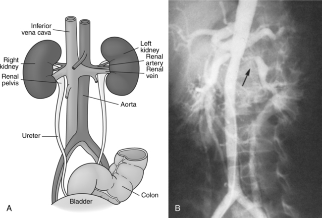
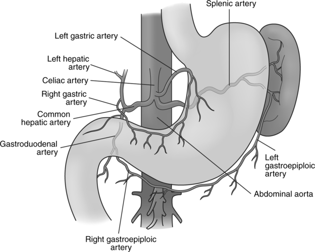
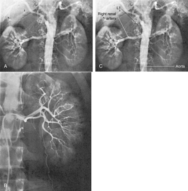
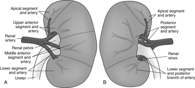
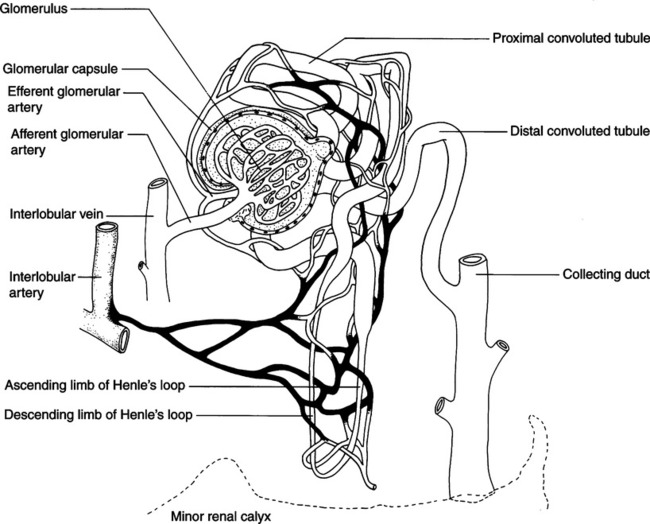
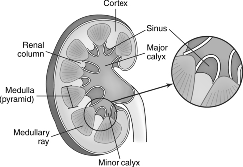
Abdominal Aortography and Genitourinary System Procedures

CHAPTER 13


Abdominal Aortography and Genitourinary System Procedures


The abdominal portion of the descending aorta begins as it passes through the diaphragm and enters the abdominal cavity at approximately the level of the twelfth thoracic vertebra. Several vessels branch off the abdominal aorta to service the kidneys, intestines, and reproductive organs and to supply the diaphragm with nutrients. Conventional abdominal aortography can be performed as a “flush” study to visualize the vasculature of the vessel and to determine the presence or absence of relevant pathology. This is usually done by positioning the catheter in the lumen of the aorta at the level of T12–L1. Pelvic aortography can also be performed by placing the catheter tip approximately 2 to 3 cm (1 inch) above the aortic bifurcation. Depending on the area of interest and the suspected pathology, the catheter can be placed selectively into any of the branches to demonstrate specific vasculature.


Anatomic Considerations


Abdominal Aorta


The abdominal aorta courses toward the midline anterior to the vertebral column. At the level of the fourth lumbar vertebra, the abdominal aorta bifurcates (splits) into the right and left common iliac arteries ((Fig. 13-1), A and B). The bifurcation can be topographically located slightly inferior and to the left of the navel. This point is considered the termination of the aorta. The common iliac arteries are the major arteries of the pelvis. Each common iliac artery divides into an internal and an external iliac artery. The external iliac arteries then form the femoral arteries of the lower extremities.


Some of the branches of the abdominal aorta are paired and others are unpaired. All of the parietal vessels are paired with the exception of the median sacral artery, which branches off at the level of the aortic bifurcation and courses toward the sacrum and coccyx. The branches of the descending abdominal aorta can be classified as either parietal or visceral and may be summarized as follows:


During aortography, it is not uncommon for some of the branches to visualize. Frequently, the circulatory supply of the kidneys will be well demonstrated. The middle adrenal arteries can occasionally be seen on an abdominal aortogram, especially in cases of renal or adrenal pathology. These are paired vessels arising from the lateral wall of the aorta at a level between the celiac trunk and the superior mesenteric arteries.


Celiac Trunk and Mesenteric Circulation


The first main branch originating on the ventral aorta is the celiac trunk. The main areas supplied by this vessel are the esophagus, stomach, liver, spleen, and pancreas (Fig. 13-2). The celiac trunk bifurcates and gives off the left gastric, splenic, and the common hepatic arteries. The left gastric artery, as its name implies, runs off to the left to supply the stomach coursing along the lesser curvature and connects with the right gastric artery. The splenic artery also runs off to the left and gives off numerous branches to supply the pancreas.


The common hepatic artery courses to the right. It subsequently divides into the right gastric, gastroduodenal, and hepatic arteries. The hepatic artery runs superiorly to divide into the right and left hepatic arteries. As stated earlier the right gastric artery follows the lesser curvature of the stomach to join with the left gastric artery. The gastroduodenal artery courses posteriorly and divides into the pancreaticoduodenal artery and the right gastric epiploic, which continues along the greater curvature of the stomach to become the left gastric epiploic artery.


The superior and inferior mesenteric arteries are unpaired vessels. They are considered visceral branches of the aorta and primarily supply the small and large intestines. The superior mesenteric arises ventrally just below the origin of the celiac trunk. The inferior mesenteric artery is also ventrally located and originates just above the bifurcation of the aorta.


The capillaries of the stomach, intestine, spleen, and pancreas bring the blood into the portal vein which carries it to the capillary-like liver sinusoids. The portal vein, the large collecting vein, collects all of the venous blood from the gastrointestinal system, spleen, and pancreas. It is formed by the superior mesenteric vein and the splenic vein. Although the inferior mesenteric vein empties into one of the two other major veins in the portal system, it is considered to be one of the veins that make up the portal vein. The portal vein then courses superiorly and divides into right and left branches. These vessels then break up into interlobular and then smaller intralobular veins, which form the sinusoids. These sinusoids drain into the central vein of each liver lobule, which merges into the hepatic veins, which drain into the inferior vena cava.


Urinary System


The kidneys are bean-shaped organs that lie in the abdomen next to the vertebral column at the level of the twelfth thoracic vertebra to the second or third lumbar vertebra. The renal hilum, an indented area on the medial border of the kidneys, can usually be found at the level of the interspace of the first and second lumbar vertebrae. This level can vary by as much as one vertebral body in either direction. It is here that the major vessels and ureters enter and leave the kidney.


The internal structure of the kidney is divided into two areas—the cortex, or outer area, and the medulla, or inner portion. The cortex contains the glomeruli, which are capsules that enclose a convoluted group of capillaries. The glomeruli are portions of the nephrons, the functional units of the kidney (Fig. 13-3). The cortex also comprises other portions of the nephron, the convoluted tubules, and portions of the origins of the collecting ducts.


The medulla comprises straight tubules, continuations of the convoluted tubules called Henle’s loop, and collecting ducts. The medulla has the appearance of many pyramids, the apex of which terminates in the cuplike indentation of the minor renal calyx (Fig. 13-4).


The nephrons filter blood plasma and permit selective reabsorption of water and dissolved materials necessary for maintaining the ionic balance of the blood back into the circulation. There are 1 million or more of these nephrons in each kidney. During renal arteriography, the interlobular arteries are not outlined as separate channels but instead appear as a diffuse accumulation over the entire kidney. This is usually called the nephrogram phase of the arteriogram.


Adrenal Gland Vasculature


The adrenal glands sit on top of each kidney. They are embedded in the adipose tissue that surrounds the kidney and consists of a cortex (outer portion) and the medulla (inner portion). Both of these areas are well supplied with blood vessels.


The medullary portion of the adrenal glands is responsible for the secretion of epinephrine (adrenaline) and norepinephrine (noradrenalin). Tumors in the adrenal glands can affect the amount of hormone that is secreted. Norepinephrine is usually increased, causing a variety of different bodily responses such as high blood pressure and increased heart rate.


The adrenal cortex also gives off a number of steroids and hormones. Three of these secretions are important for the proper maintenance of electrolyte balance and glucose metabolism: (1) the adrenal sex hormones—adrenal androgen in the male and its converted form, estrogen, in the female; (2) aldosterone; and (3) cortisol (hydrocortisone). Unlike the secretions from the medulla of adrenal medulla, if the cortical secretions are interrupted for even a week, survival of the patient is questionable.


The adrenal glands are supplied with blood via three arteries to each gland: the superior, middle, and inferior adrenal arteries. The superior adrenal artery is a branch of the inferior phrenic artery. The middle adrenal is a direct branch from the aorta, arising at some point between the celiac and the renal arteries. The inferior adrenal artery branches off each of the renal arteries. These vessels break into many smaller branches that infuse both the cortical and medullary portions of the gland. As the vessels pass through the medulla they become smaller capillaries that begin to take on the characteristics of venules.


When they approach the center of the medullary area they join into a central vein. The venous vasculature differs between the left and right adrenal gland. In the right adrenal gland there are generally three major branches coming from the posterior, superior, and inferior portions of the gland. These join into a larger branch which communicates with the inferior vena cava.


The left adrenal gland has a single central vein that joins the left phrenic vein just before emptying into the left renal vein.


Renal Circulation


The renal arteries originate as branches of the abdominal aorta at about the level of the interspace of the first and second lumbar vertebrae (Fig. 13-5). They run transversely toward each kidney, with the right renal artery passing posterior to the inferior vena cava. The superior mesenteric artery is just above the origin of the renal arteries.


The renal artery gives off an extrarenal branch called the inferior suprarenal artery, which supplies the largest portion of the adrenal gland. Other portions are supplied by the superior and middle suprarenal arteries, which are branches originating directly from the aorta. The adrenal glands are usually demonstrated during renal angiography.


Before the renal arteries enter the hilum of the kidney, each divides into five branches corresponding to the five renal segments. These enter the renal sinus and course both anteriorly and posteriorly to the renal pelvis (Fig. 13-6). The renal sinus is a recess containing the renal vessels and the renal pelvis, the upper expanded portion of the ureter.


In the renal sinus, the larger branches of the renal artery subdivide to form smaller interlobar branches. These vessels course between the lobes, in the renal columns, toward the periphery of the kidney. At the level of the renal cortex, the interlobar branches become the arcuate arteries. These arteries course along the line between the cortex and medulla and give off branches called the interlobular arteries.


The interlobular arteries subdivide into the afferent arterioles that course into the glomerulus. In the glomerulus, the afferent arteriole becomes an efferent arteriole and then forms a capillary plexus surrounding the straight and convoluted tubules.


The efferent arteriole plexus is continuous with small venules that unite to form interlobular and medullary veins. These drain into the arcuate veins, which course along the boundary of the cortex and medulla. Interlobar veins are formed by the arcuate veins and converge to form the renal vein, which ultimately drains into the inferior vena cava.


Table 13-1 summarizes, in order of filling, the vessels seen during the various phases of renal arteriography. It should be noted that when renal masses are evaluated with intravenous digital subtraction angiography (DSA), the catheter is placed in the inferior vena cava via the femoral approach. This technique demonstrates the patency of the inferior vena cava during the initial phase of the study. The later phases appear as stated in Table 13-1


Feb 27, 2016 | Posted by in GENERAL RADIOLOGY | Comments Off on Abdominal Aortography and Genitourinary System Procedures

Full access? Get Clinical Tree

Get Clinical Tree app for offline access