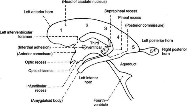
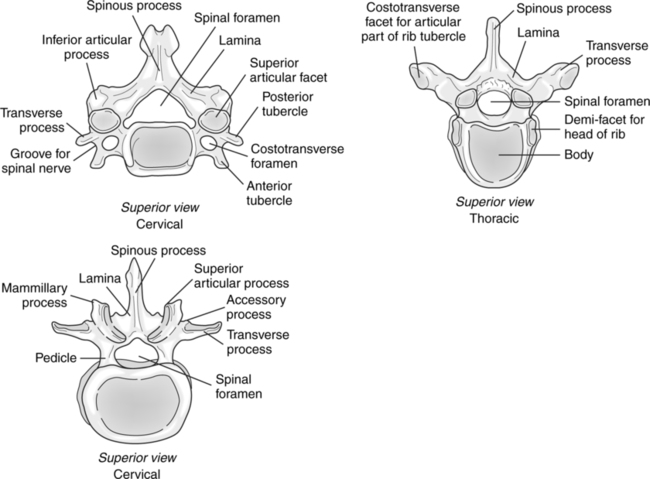
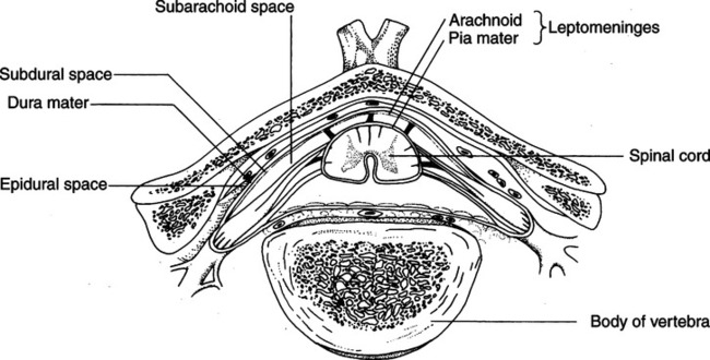
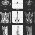
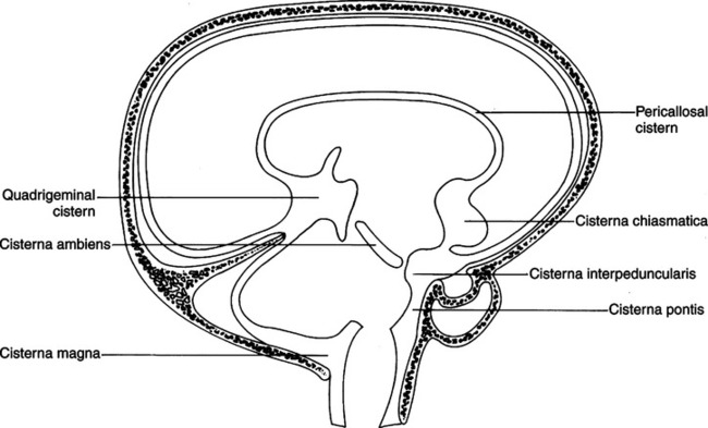
Myelography

CHAPTER 21


Myelography


Radiography of the structures of the central nervous system has undergone a number of major changes in recent years. Newer modalities such as computed tomography (CT) and magnetic resonance imaging (MRI) have completely replaced invasive studies such as pneumoencephalography, ventriculography, and, to some extent, myelography.


Myelography is the radiographic demonstration of the central nervous system structures located within the spinal canal. The procedure is accomplished easily through direct instillation of a contrast agent into the subarachnoid space. This technique can demonstrate various abnormalities of the spinal cord and spinal canal.


ANATOMIC CONSIDERATIONS


The nervous system can be separated into two major divisions—the peripheral and the central nervous systems. The central nervous system consists of the brain and the spinal cord. The central nervous system processes information to and from the peripheral nervous system and is the control center for the body.


Brain


The brain is the portion of the central nervous system found in the cranial cavity. The brain comprises the pons, medulla, mesencephalon, diencephalon, cerebellum, and cerebrum.


The pons, medulla, mesencephalon, and diencephalon are the components of the brain stem. The function of the brain stem is to provide some motor, sensory, and reflex functions. The spinal cord extends from the medulla oblongata and is considered to begin at the level of the foramen magnum.


The cerebellum is also called the hindbrain and is located in the posterior cranial fossa behind the brain stem. The cerebellum consists of two hemispheres separated by a groove. The middle section of the cerebellum, the vermis, is the connection between its hemispheres. The main function of the cerebellum is to coordinate voluntary muscular activity.


The cerebrum is the largest and uppermost portion of the brain. It is divided into right and left hemispheres by a central groove, the sulcus, and is connected at the bottom of the groove by the corpus callosum. The surface of the cerebrum is convoluted and lobed. The cerebrum is composed of an outer cerebral cortex (gray matter) and an inner portion, or semiovale (white matter). The cerebrum provides the sensory, motor, and integrative functions associated with the body’s mental and physical activities. This portion of the brain generates the electric waves that are monitored and recorded with an electroencephalograph.


Subarachnoid Cisternae


In some areas of the brain, the subarachnoid space enlarges to form the subarachnoid cisternae (Fig. 21-1). The cisterna magna extends down to merge with the spinal subarachnoid space. It is triangular and contains approximately 5 to 10 ml of cerebrospinal fluid. The apex of the cisterna magna points toward the vallecula. This is the portion of the subarachnoid space that lies between the fourth ventricle and cisterna magna. When air is injected into the spinal subarachnoid space it must pass through the cisterna magna and vallecula before entering the ventricular system. The flexion of the head must be accurately maintained to prevent air from entering other subarachnoid spaces rather than the ventricular system.


The ventricular system is a series of spaces or cavities located within the hemispheres of the brain. The system consists of four cerebral ventricles—two lateral ventricles and two other ventricles called the third and the fourth ventricles. Cerebrospinal fluid circulates in the ventricles and subarachnoid space surrounding the brain and spinal cord. The term “circulate” when applied to cerebrospinal fluid should not be compared with “circulation” as it applies to the blood. Cerebrospinal fluid is a complex substance containing various components, including water, electrolytes, and proteins. These substances are produced or absorbed in varying amounts throughout the subarachnoid space. This is the basis for the circulation of the spinal fluid. The cerebrospinal fluid flows from areas of greatest production to areas of greatest absorption, but it is not true circulation. However, the spinal fluid does move through the subarachnoid system by this production-absorption process.


Lateral Ventricles.

There are two lateral ventricles, each located within one cerebral hemisphere. Each lateral ventricle has five divisions (Fig. 21-2): (1) the anterior (frontal) horn; (2) the body; (3) the trigone (isthmus or atrium); (4) the posterior (occipital) horn; and (5) the inferior (temporal) horn. Each lateral ventricle connects on each side with the third ventricle by a narrow channel known as the interventricular foramen of Monro. The anterior (frontal) horns are usually found in the frontal lobes of the brain hemispheres.


Spinal Cord


The vertebral canal tends to be triangular, relatively large in the cervical and lumbar regions, and small and ovoid in the thoracic region (Fig. 21-3). The structures contained in the vertebral canal are the spinal cord and its meninges, spinal nerves and vessels, and the epidural space, which is located between the wall of the vertebral canal and the dura mater. The epidural space contains fat, venous plexuses, and nerves that supply the meninges, intervertebral disks, and ligaments.


All of these structures are important in that any aberrations may cause encroachment on the vertebral canal. The spinal cord lies loosely within the vertebral canal and extends from the foramen magnum to the lower border of the first lumbar vertebra. At this point, the spinal cord tapers into the conus medullaris, from which the filum terminale extends to the coccyx. The spinal cord averages 45cm (18 in) in length.


The three layers—the dura mater, or outer covering; the arachnoid, middle layer; and the pia mater, the inner layer—also enclose the spinal cord and are continuous with the layers surrounding the brain (Fig. 21-4).


The spinal dura mater is a heavy sheath extending from its attachments with the margins of the foramen magnum to the level of the second sacral vertebra. At this point, the dura mater tapers into a covering for the filum terminale of the spinal cord. A space exists between the wall of the vertebral canal and the dura mater; this is called the epidural space. This space contains semifluid fat and many small veins. Between the dura mater and arachnoid layers is a potential subdural space. The meningeal layers contact each other, with a film of fluid separating them.


The spinal arachnoid is continuous with the cerebral arachnoid layer that covers the brain. It is a delicate membrane that follows the dura mater to its termination at the second sacral vertebra. Between the arachnoid and pia mater is the subarachnoid space. This space is bathed in spinal fluid and is in direct communication with the ventricles of the brain and its surrounding spaces. The subarachnoid space is larger around the spinal cord than in the brain. It extends to the level of the second sacral vertebra. The spinal cord ends at the upper level of the lumbar spine and the subarachnoid space continues to the second sacral segment, making an ideal location for spinal fluid withdrawal or contrast medium injection. The nerve roots are seldom damaged because of their flexibility in this area.


The spinal pia mater is the innermost membrane that invests the spinal cord. It is a thin layer that contains some blood vessels. The pia mater extends into the filum terminale and terminates within it, blending into the periosteum at the posterior of the first coccygeal segment. The pia mater and arachnoid are occasionally referred to as one layer. When this is the case, they are called the pia arachnoid layer, or the leptomeninges.


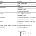
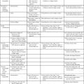
INDICATIONS AND CONTRAINDICATIONS


Subarachnoid myelography is a safe procedure that can be used to visualize the subarachnoid space around the spinal cord. It is indicated when accurate diagnosis of anatomic abnormalities or pathologic processes of the spinal cord and spinal canal is required. Some specific indications for subarachnoid myelography are as follows:


Epidural myelography is indicated when a demonstration of encroachment defects caused by tumors or herniated intervertebral disks into the lower thoracic and lumbar regions of the spinal canal is necessary.


Myelography is contraindicated when the patient exhibits signs of increased intracranial pressure.


PATIENT PREPARATION


In many cases, myelography can be done as an ambulatory procedure. When the examination is scheduled, patients should be instructed not to take medication for at least 24 hours and to increase fluid intake before the examination. It is also recommended that they not eat solid food before the study; the length of the fast is variable depending on the protocol of the institution.


When the patient arrives, the procedure should be explained and a consent form negotiated. The patient should be questioned concerning allergies, prior contrast agent reactions, results of any recent myelograms, and history of seizures. Vital signs should be taken at this time to establish a baseline for monitoring during the procedure. The information from the history should be transmitted to the physician before the study begins. If the patient is to receive any medication before the examination, it should be administered by the physician or the special procedure nurse.


The radiographic room should be set up before the patient is brought in. All equipment should be tested, and the supplies necessary for the injection should be placed within easy reach. The patient should be gowned in preparation for the study. The injection site should be shaved; when the physician arrives, the site should be surgically prepared.


During the procedure, the patient should be monitored for changes and any unusual symptoms or complaints. If specimen samples are required, care must be taken to preserve the sterile environment. Samples should be readied for laboratory analysis as soon as possible. The procedure will be determined by the protocols developed by the medical laboratory department and must be carefully followed. Any complications or reactions should be noted on the patient’s chart; the individual responsible for the charting will vary depending on the hospital protocol.

Stay updated, free articles. Join our Telegram channel

Feb 27, 2016 | Posted by in GENERAL RADIOLOGY | Comments Off on Myelography

Full access? Get Clinical Tree

Get Clinical Tree app for offline access