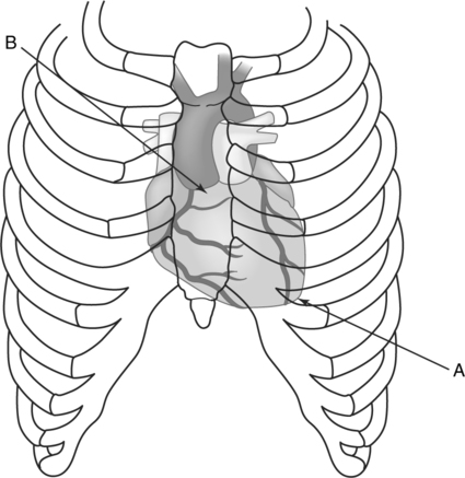
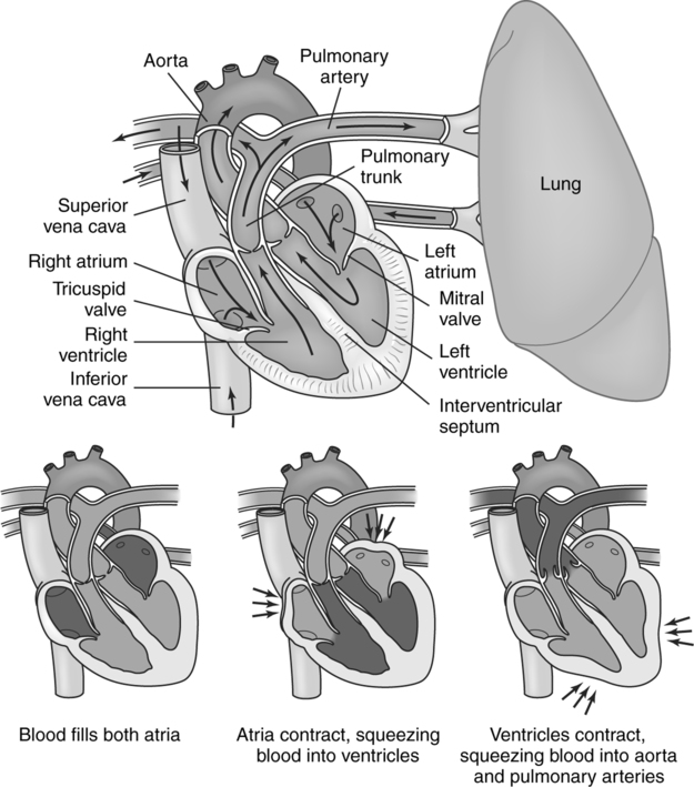
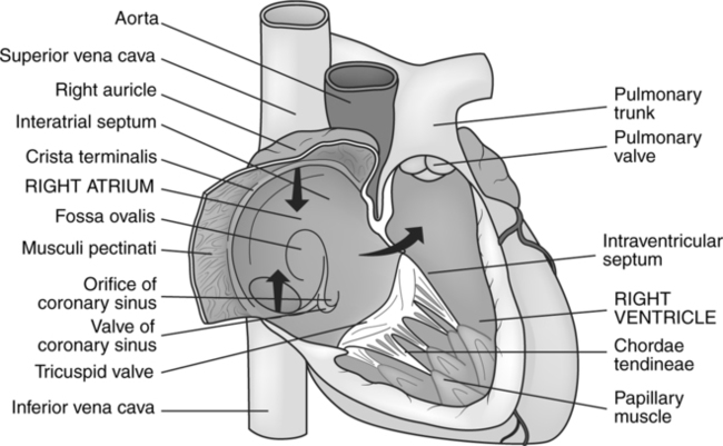
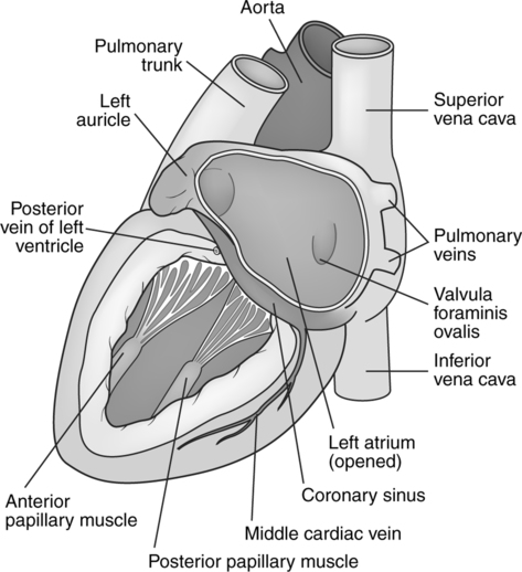
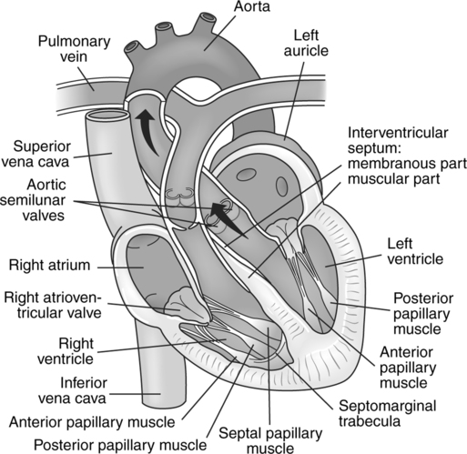
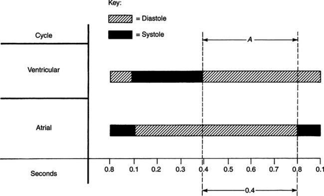
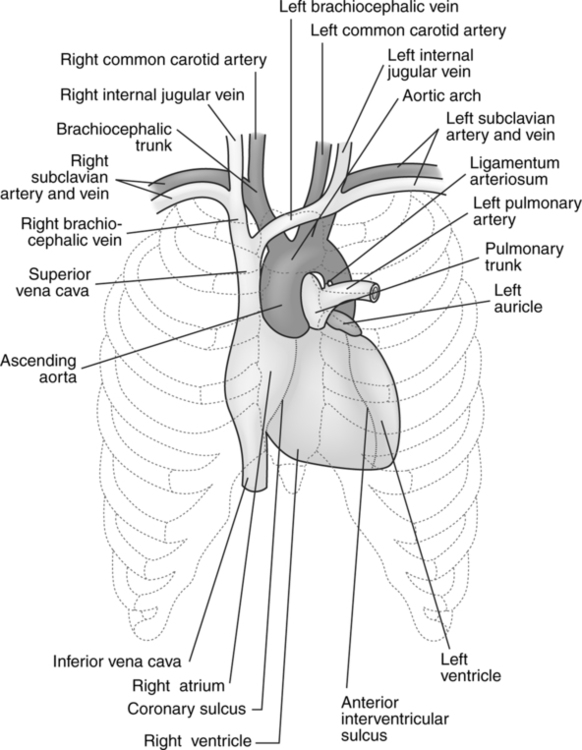
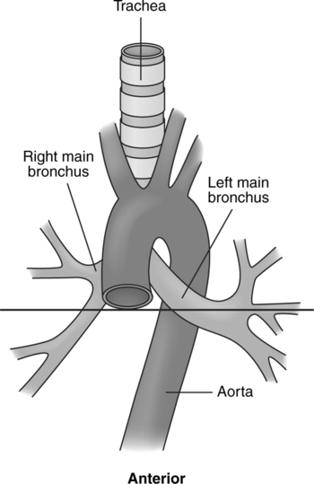
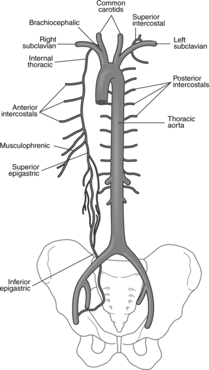
Cardiac and Thoracic Procedures

CHAPTER 12


Cardiac and Thoracic Procedures


Numerous diagnostic studies can be performed on the vasculature of the heart, lungs, and thoracic aorta. In most cases these diagnostic procedures are done with the intent of performing interventions, if possible, in order to correct any pathophysiology that might be identified. The common cardiac and vascular interventions that can be performed will be discussed in Chapters 17 and 18. The interventional studies usually involve the same parameters as the diagnostic procedures, and the basic principles from the diagnostic chapters will not be repeated in the discussion about interventions.


Generalized vascular pathology will be discussed in this chapter. The pathophysiology of the vasculature spans the entire circulatory system. The following discussion will serve to lay the groundwork for the various disease processes that will be treated in the interventional section of this book.


The thoracic cavity contains the heart, great vessels, aorta, and pulmonary circulation. Some of the procedures that are performed in this area are aortography, cardiac catheterization, coronary arteriography, and pulmonary arteriography. Discussion of the studies performed on the abdominal aorta and its associated vessels can be found in Chapters 13 and 14.


The circulatory system is responsible for moving the blood throughout the body in order to bring oxygen and nutrients to the various tissues and remove any waste products for elimination. It consists of a pump (the heart), conduits to move the blood from place to place (arteries and veins), and locations where the nutrients and wastes are transferred (lungs, kidneys, and gastrointestinal system). As in any closed system, problems can occur that compromise its normal function. The common situations that cause malfunction of the system are leaks, reduced or increased flow, and directional anomalies that can cause flow in the wrong direction. The purpose of diagnostic and interventional angiography is to identify and possibly correct these problems.


Before a discussion of the gross anatomy of the circulatory system can be held it is important to understand the structure of the blood vessels, one of the important components of the circulatory system. The gross anatomy of the heart, location of the blood vessels, and operations of the transfer stations (lungs, kidneys, and gastrointestinal system) will be discussed in each of their representative chapters.


NORMAL BLOOD VESSEL STRUCTURE


Normal blood vessels are muscular tubes that consist of three layers: the tunica intima (interna), tunica media, and tunica adventitia (externa). The tunica intima is essentially a lining of endothelial cells over a layer of connective tissue separating the blood from the vessel proper. This layer is important for maintaining homeostasis in the circulatory system and subsequently is the site of much of the vascular pathology. It gets its nutrients from direct diffusion from the lumen of the vessel. Small arterioles (vasa vasorum) that permeate the adventitia and media provide the vessels with a supply of nutrients and oxygen.


The endothelial cells secrete a variety of hormones that allow the vessel to react to a variety of stressors and are vital in the vessel’s response to injury. The endothelial cells also help to prevent clotting by secreting chemicals that inhibit the aggregation of platelets. Substances are also secreted by the endothelial cells that help control the relaxation (vasodilation) and contraction (vasoconstriction) of the vessels.


The media is the next layer of the blood vessels. This layer is composed of smooth muscle cells, elastic fibers, and connective tissue. The muscle cells and connective tissue of this layer provide support for the vessel walls, while the elastic fibers give it the capability to respond to the pressure changes from the systolic and diastolic action of the heart by changing the diameter of the vessel. Arteries have a larger muscular layer than do veins because they are subject to the direct pressures from the contractions and relaxation phases of the heart. The movement of blood in the veins is controlled by pressure gradients, folds in the endothelial layer that act in a similar manner to the valves of the heart (preventing the backflow of blood) and the contraction of the muscles surrounding the veins. The amount of elasticity depends upon the vessel. Larger arteries such as the aorta have a greater response than do smaller less elastic arteries.


The outer layer of the vessels is composed primarily of a thin layer of connective tissue, irregular elastic, and collagen fibers. This is the means by which the vessels are attached to the local body tissues. The tunica adventitia (externa) of the larger veins and their tributaries has a thicker layer of collagen and elastic fibers than do the arteries. Overall, the veins have relatively thin walls when compared to the arteries because they do not have to withstand the types of pressure changes that occur in the arteries. It is because of this factor that the veins do not have a similar pathophysiology to the arteries.


The arteries and veins of the circulatory system are connected by means of the capillary bed. As the vessels get smaller and smaller they form many branches that create the capillary network. The arteries become arterioles (very small vessels), which give off many smaller vessels called capillaries. These capillaries then collect into a group of venules (very small vessels that resemble the arterial capillaries). The capillary beds are where the diffusion of materials takes place across the walls of these small vessels.


COMMON VASCULAR PATHOPHYSIOLOGY


The arteries are subjected to a number of pathologic situations because of their structure and function. The endothelial lining of the vessel is designed to provide a smooth surface for the blood cells and platelets to move easily through the lumen. Just as in home plumbing, the action of the materials passing through the pipes can cause a narrowing of its inside diameter, restricting the flow of water. The arteries are similar in that some of the materials (platelets) passing through the vessel may become lodged on the interior wall of the vessel, and the prolonged exposure to the high pressures from the heart, trauma, or infection can trigger a response from the cells of the intima. The normal physiology of the cells of the tunica intima can cause the deposition of new endothelial cells over the area, reducing the vessel lumen and ultimately the flow of blood. The narrowing can be the result of arterial stenosis (atherosclerosis), thrombosis, or intimal hyperplasia. Stenosis is also found in veins, although not as often because the pressures are lower in the system.


Blockage of the vessel can occur if some material (embolus) dislodges and travels through the circulatory path until it becomes lodged in one of the vessels. The blockage effectively stops the flow of blood to the distal vessels and over time the blockage increases on the proximal side of the vessel. This type of pathophysiology lends itself to treatment utilizing interventional techniques.


Another common pathophysiologic process results from the constant expansion and contraction of the artery due to the pulsations from the heart. Exposure to a high level of pressure change can cause the vessel to lose its elasticity, causing an abnormal enlargement of the vessel (greater than 50% of its original diameter) accompanied by a thinning of the media and adventitia due to the loss of collagen, muscle, and elastic fibers. Veins, on the other hand, are not as prone to aneurysms (abnormal enlargement with vessel weakness) as arteries; however, they can lose the capacity for normal valve action, creating a condition of reversed blood flow.


Various congenital arteriovenous malformations can result in the increased flow of blood. These are generally present at birth and can increase in size and complexity as the child develops, making interventional treatment difficult. Malformations can occur in the venous system as well; however, these defects are not high flow disorders and are easier to treat by means of interventional radiographic procedures.


ANATOMIC CONSIDERATIONS
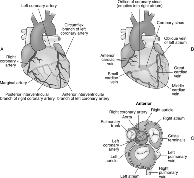

Heart


The heart is a hollow muscular organ that lies obliquely in the left median portion of the lower thoracic cavity. It adjusts the circulation of the blood to the metabolic rate of the tissue cells; in essence, it is a pump.


The heart has an apex and a base. The apex is located approximately 8 cm from the median plane at about the level of the fifth or sixth interspace; this level is approximately 2.5 cm inferior to the left nipple. The base of the heart is the most superior portion. It faces up and to the right, and from it emerge the greater vessels (Fig. 12-1).


An obliquely placed septum divides the heart into right and left halves. Each half consists of two chambers—an atrium and a ventricle (Fig. 12-2).


The atria receive blood from the veins, and the ventricles propel blood into the arteries. Blood from the heart flows in two circuits—a short circulation (lesser circuit) called the pulmonary and a longer, more extensive one (greater circuit) called the systemic.


The anatomy of the heart is easily understood if it is related to the flow of blood through it. Therefore, the anatomy of the heart is presented in relation to the blood flow, beginning with the drainage from the systemic circulation.


Blood returning from the systemic veins drains into the venae cavae. These great veins—the superior and inferior venae cavae—empty into the right atrium of the heart (Fig. 12-3). A small amount of venous blood drains from the myocardium directly into the right atrium; however, in general, the greatest portion of venous blood from the systemic circulation drains into the superior and inferior venae cavae.


With the contraction (systole) of the right atrium, the blood is forced into the right ventricle through the right atrioventricular aperture. This aperture is equipped with a valve, the tricuspid valve, that prevents the backflow of blood to the right atrium during systole of the ventricle. As the ventricle contracts, the blood is forced past the pulmonary semilunar valve into the pulmonary trunk and the lesser circulation. The arterial, or outflowing, portion of the right ventricle is called the conus arteriosus.1 The pulmonary artery divides into right and left sides, which take the blood through the lungs. Here, the blood gives off carbon dioxide and becomes oxygenated. It is then transported by the four pulmonary veins to the left atrium (Fig. 12-4).


As the left atrium contracts, it forces the blood through the left atrioventricular valve into the left ventricle. This is the largest chamber of the heart, and its walls are considerably thicker than those of any other chambers. The thicker walls and larger capacity are necessary to offset the arterial pressure in the aorta. The upper anterior portion of the left ventricle is called the aortic vestibule.1 As the left ventricle contracts, the blood is pushed through the aortic semilunar valve into the aorta and through the systemic circulation (Fig. 12-5). The blood returns from the body tissues through the systemic veins, which ultimately drain into the superior and inferior venae cavae; then the cycle begins again.


The cardiac cycle is repeated about 75 times per minute, and the time required for each cycle is about 0.8 s. The cycle consists of three phases—atrial contraction(systole), ventricular contraction (systole), and complete rest (diastole) (Fig. 12-6).


Thoracic Aorta


The aorta can be subdivided into four parts — the aortic root, ascending aorta, aortic arch, and descending aorta.


The aortic root or bulb begins at the level of the aortic semilunar valve. The three leaflets of the aortic valve form the aortic sinuses, right, left, and posterior. The right and left aortic sinus house the openings to the coronary arteries, which provide the heart muscle with the necessary nutrients for survival.


The ascending aorta originates at the base of the heart with the root of the aorta. The aortic sinuses are found in the wall of the root of the aorta. These are related to the cusps of the aortic valve and are named for them—right, left, and posterior. Two of these sinuses—the right and the left—contain the orifices of the coronary arteries. The ascending aorta extends anterosuperior from the base of the heart and slightly to the right for approximately 5 cm and terminates by becoming the arch of the aorta, which is usually at the level of the sternal angle (Fig. 12-7).


The aortic arch courses from right to left as well as from anterior to posterior. It lies in almost a true sagittal plane (Fig. 12-8). From the sternal angle, the aortic arch ascends toward the left. As it ascends, it is directed posterior. The upper portion of the arch courses posterior to the left of the trachea and esophagus. At about the level of the fourth thoracic vertebra, it turns inferior and runs a short distance before becoming the descending aorta.


There are three major branches given off by the aortic arch—the brachiocephalic trunk, the left common carotid artery, and the left subclavian artery (Fig. 12-9). These branches supply the head and upper extremities with blood.


The descending aorta is a continuation of the aortic arch. It can be divided into a superior thoracic descending and an inferior abdominal descending portion. The thoracic descending aorta extends from its origin at the level of the intervertebral disk between the fourth and fifth thoracic vertebrae to the point at which it traverses the aortic hiatus in the diaphragm (Fig. 12-10), which is about the level of the twelfth thoracic vertebra. As the thoracic aorta passes through the opening in the diaphragm, it becomes the abdominal portion of the descending aorta.


The branches of the thoracic descending aorta can be classified as either parietal or visceral and may be summarized as follows:


The parietal branches supply the body wall of the thoracic cavity, whereas the visceral branches supply the organs contained within the thoracic cavity.


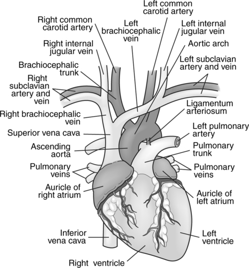
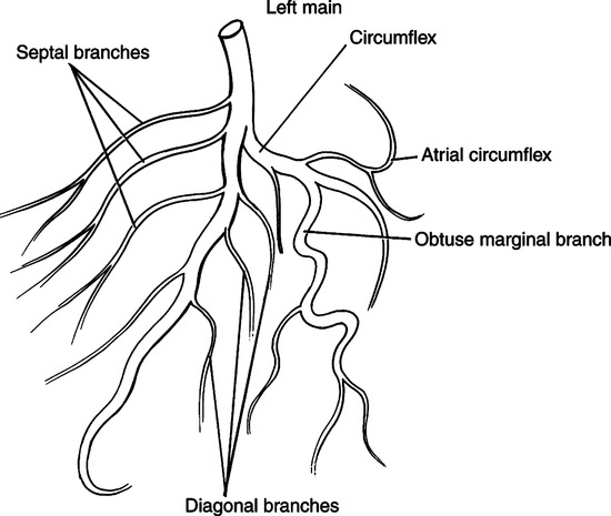
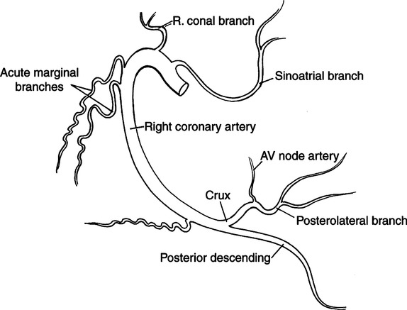
Coronary Vasculature


The coronary arteries are the source of blood supplying the heart. The aorta is the origin of the two main coronary arteries (Fig. 12-11). Three small dilations, or sinuses, are located in the root of the aorta. They lie opposite the corresponding cusps of the aortic valve. The coronary arteries arise from two of these, so they are considered coronary sinuses. The third sinus is considered a noncoronary sinus because it does not connect directly with any coronary arteries.


The left main coronary artery arises from the left posterior aortic sinus, and the right main coronary artery originates from the anterior, or right, aortic sinus. These sinuses are called the aortic sinuses, or Valsalva’s sinuses.


Left Main Coronary Artery


This artery originates in the left aortic sinus. It arises from a single opening in the upper portion of the sinus, and its length can vary from 2 to 3 mm to 3 to 4 cm. It divides to form the anterior interventricular branch (left anterior descending artery) and the left circumflex branch (Fig. 12-12). These branches occupy the anterior interventricular sulcus and the atrioventricular sulcus, respectively. This oversimplification of the bifurcation of the left main coronary artery is provided because usually no more than two large branches of this artery can be found.1


The anterior interventricular branch is considered a direct continuation of the left main coronary artery. It courses toward the apex of the heart in the anterior interventricular sulcus. In the frontal view, the left anterior interventricular branch plus the left main coronary artery form a gently reversed “S” curve. After reaching the apex of the heart, the left anterior interventricular branch continues up into the posterior interventricular sulcus and anastomoses with the terminal branches of the posterior interventricular branch of the right coronary artery. Along its route, the left anterior interventricular branch gives off a variable number of arteries that course into the interventricular septum. At the level of the pulmonary valve, smaller branches are given off that supply the right ventricle. One or more of these branches form a curve around the heart and meet similar branches of the proximal right coronary artery. This circle is called Vieussens’ ring, and it is an important means of collateral circulation between the right and left coronary arteries. Some larger branches are given off to supply the free wall of the left ventricle. At times, one or more of these branches arise at the point of division of the left main coronary artery; these are usually called the diagonal left ventricular arteries.


The left circumflex coronary artery, which is the second major branch of the left main coronary artery, courses at a sharp angle from its origin toward the left atrioventricular sulcus. It travels in this groove around to the back of the heart toward the right coronary artery, where anastomosis of these arteries frequently occurs. The circumflex artery gives off a large branch called the left marginal artery. The left marginal artery courses along the left margin of the heart to supply the left ventricle. Branches of the left marginal artery course over the free wall of the left ventricle somewhat parallel to those given off by the left anterior interventricular branch (left anterior descending artery).


Right Main Coronary Artery


Frequently, the right main coronary artery has two aortic ostia, which may arise from the anterior aortic sinus and from a smaller adjacent ostium. When present, the second ostium is very small—about 1 mm in diameter. This is called the conus artery of the right coronary system (Fig. 12-13). It is this artery that frequently anastomoses with an opposing branch from the left anterior interventricular artery, forming Vieussens’ ring. When the right main coronary artery arises from a single ostium in the aortic sinus, the first ventricular branch is considered the conus artery.


From its origin in the aortic sinus, the right main coronary artery courses in the right atrioventricular sulcus (coronary sulcus) to the diaphragmatic surface of the heart. At the acute margin of the heart, the right coronary artery gives off an artery called the right marginal branch, which courses almost to the apex of the heart. On the diaphragmatic surface of the heart, the right coronary artery makes a U turn and courses toward the apex of the heart in the posterior interventricular sulcus. This terminal portion of the right coronary artery is called the posterior interventricular branch (posterior descending artery).


The U turn of the right coronary artery is located at the crux of the heart. This is the point at which the right and left atrioventricular sulci cross the posterior interatrial and interventricular sulci. It is also here that the atrioventricular node artery originates from the right coronary artery.


On reaching the apex of the heart, the posterior interventricular branch frequently anastomoses with the anterior interventricular branch of the left coronary artery.


Venous Drainage


The venous drainage of the heart is considered to consist of three separate groups, rather than one.


The first group from the area of the left ventricle drains into the coronary sinus. Some prominent veins in this system are the anterior interventricular, posterior interventricular (middle cardiac), and left marginal veins. As it enters the atrioventricular sulcus, the anterior interventricular vein becomes the great cardiac vein, which in turn becomes the coronary sinus. The posterior interventricular vein usually drains directly into the right atrium; however, it often joins the coronary sinus just before entering the atrium. The left marginal vein and associated smaller vessels drain from the left ventricle into the great cardiac vein and coronary sinus.


In the second group, several large veins called the anterior cardiac veins transport venous blood from the area of the right ventricle and drain directly into the right atrium. The most prominent of these veins—the right marginal vein—drains the lower margin of the heart.


The third group consists of many very small veins, thebesian veins, which are often found on the right side of the heart but seldom on the left side. These small venous tributaries begin in the myocardium and drain directly into the cardiac chambers. They are found primarily in the right atrium and right ventricle.


Pulmonary Circulation


The right portion of the heart receives oxygen-poor blood via the superior and inferior venae cavae and directs it to the pulmonary circulation for oxygenation. Some blood is also sent to the bronchial arteries to supply the trachea, bronchi, esophagus, and posterior mediastinum. The pulmonary vasculature is also supplied with nutrients by the small vessels called the vasa vasorum. The pulmonary circulation can be divided into three portions: the pulmonary arteries, the alveolar capillary system, and the pulmonary veins. The right atrium propels the blood through the main pulmonary artery (pulmonary trunk), which branches off into a right and left main pulmonary artery. Each of these arteries services its respective lung, entering at the level of the hilum.


Left Pulmonary Artery


As the left pulmonary artery enters the hilum it gives off two branches corresponding to the upper and lower lobes of the lung. These branches also subdivide, corresponding to the bronchopulmonary segments, and terminate in the alveolar capillary beds.


The capillary beds in the alveoli are where the gaseous exchange of oxygen and carbon dioxide from the blood to the air occurs.


The blood moves through the pulmonary capillary beds into the small venules. These unite into larger and larger channels, maintaining the same association with the bronchopulmonary segments that the arteries demonstrated. This continues until a single vein for each of the lobes of the lung is formed. The vein from the middle lobe and superior lobe of the right lung join, resulting in two pulmonary veins leaving the right lung. The veins exiting from the left lung also result in two vessels. The four pulmonary veins end in the left atrium of the heart. From here the blood is passed through to the left ventricle and out into the systemic circulation.


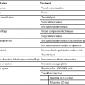
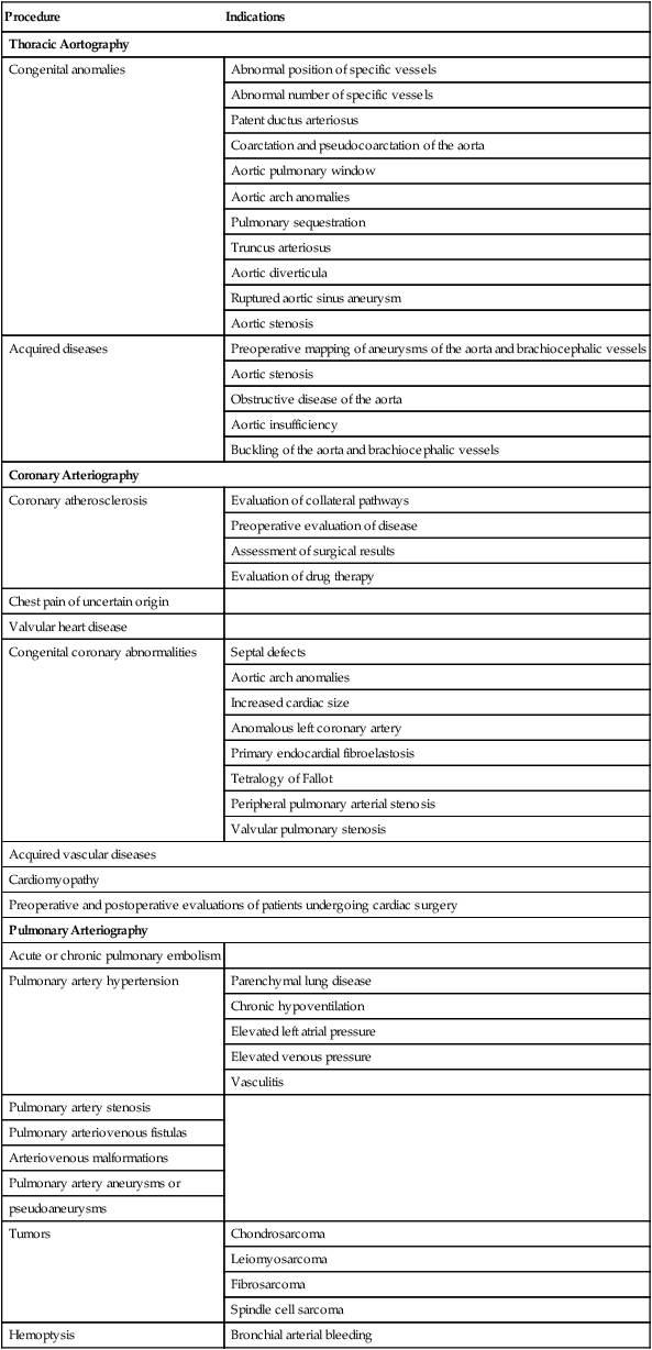
INDICATIONS AND CONTRAINDICATIONS


Aortography is indicated when information concerning the state of the aorta and its branches is necessary for therapeutic, surgical, or endovascular intervention. However, the procedure has been all but replaced as a screening tool for diagnosis by magnetic resonance angiography (MRA) and three-dimensional computed tomography (3D CT). Nevertheless, catheter aortography is still used as a preoperative tool by many surgeons because it allows an accurate preoperative evaluation concerning the nature, number, and course of the great vessels of the chest and abdomen. As they become more refined, MRA and 3D CT will more than likely replace catheter aortography as the method of choice for evaluation of the aorta and its branches.


Some specific indications for the procedure include suspected aneurysms, congenital anomalies, and many acquired diseases affecting the thoracic aorta. Table 12-1 lists specific indications for angiography of the thoracic aorta, cardiac circulation, and pulmonary circulation.


TABLE 12-1


Summary of Indications for Angiography of the Structures in the Thoracic Cavity


Procedure Indications
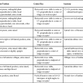
Thoracic Aortography
Congenital anomalies Abnormal position of specific vessels
Abnormal number of specific vessels
Patent ductus arteriosus
Coarctation and pseudocoarctation of the aorta
Aortic pulmonary window
Aortic arch anomalies
Pulmonary sequestration
Truncus arteriosus
Aortic diverticula
Ruptured aortic sinus aneurysm
Aortic stenosis
Acquired diseases Preoperative mapping of aneurysms of the aorta and brachiocephalic vessels
Aortic stenosis
Obstructive disease of the aorta
Aortic insufficiency
Buckling of the aorta and brachiocephalic vessels
Coronary Arteriography
Coronary atherosclerosis Evaluation of collateral pathways
Preoperative evaluation of disease
Assessment of surgical results
Evaluation of drug therapy
Chest pain of uncertain origin  
Valvular heart disease  
Congenital coronary abnormalities Septal defects
Aortic arch anomalies
Increased cardiac size
Anomalous left coronary artery
Primary endocardial fibroelastosis
Tetralogy of Fallot
Peripheral pulmonary arterial stenosis
Valvular pulmonary stenosis
Acquired vascular diseases
Cardiomyopathy
Preoperative and postoperative evaluations of patients undergoing cardiac surgery
Pulmonary Arteriography
Acute or chronic pulmonary embolism  
Pulmonary artery hypertension Parenchymal lung disease
Chronic hypoventilation
Elevated left atrial pressure
Elevated venous pressure
Vasculitis
Pulmonary artery stenosis  
Pulmonary arteriovenous fistulas
Arteriovenous malformations
Pulmonary artery aneurysms or
pseudoaneurysms
Tumors Chondrosarcoma
Leiomyosarcoma
Fibrosarcoma
Spindle cell sarcoma
Hemoptysis Bronchial arterial bleeding


image


Thoracic aortography and cardiac catheterization are accomplished primarily to demonstrate the coronary arteries and cardiac anatomy as well as to perform hemodynamic measurements to identify a variety of disease processes. The studies can be done to demonstrate pathophysiology of the thoracic aorta and pulmonary and peripheral vasculature. Interventions can also be accomplished as an adjunct to the diagnostic procedures or as stand-alone procedures once the pathology has been defined.


The contraindications to angiography can be divided into those that are physiologic in nature and those that are related to the toxic effects of the contrast agents. Physiologic contraindications, which are also related to the specific procedural techniques used, include femoral arteriosclerosis, which contraindicates the retrograde transfemoral approach to avoid intimal damage, and aneurysms at the injection site, which contraindicate the use of the direct puncture technique. Some other contraindications to these procedures are infection, fever, high blood pressure, acute gastrointestinal bleeding, and renal failure

Stay updated, free articles. Join our Telegram channel

Feb 27, 2016 | Posted by in GENERAL RADIOLOGY | Comments Off on Cardiac and Thoracic Procedures

Full access? Get Clinical Tree

Get Clinical Tree app for offline access