Fig. 23.1
Diagram of congenital lesions of the urachus. (a) Patent urachus. (b) Urachal cyst. (c) Urachal sinus. (d) Vesicourachal diverticulum
23.5.2 Abscess in the Urachal Remnant
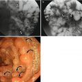
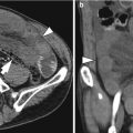
Fig. 23.2
Abscess in the urachal remnant. (a) Transverse CT image shows a small rim-enhancing mass below the anterior peritoneum. (b) Sagittal CT image shows an elongated wall-enhancing lesion (white arrowheads) from the umbilicus (black arrowhead) to the dome of bladder (asterisk), indicating that it is an abscess along the urachal remnant. After antibiotic treatment, the thickened wall was improved (not shown here)
23.5.3 Meckel’s Diverticulum in a Patient with History of Recurrent Hematochezia
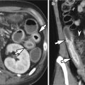
Fig. 23.3
Meckel’s diverticulum in a patient with history of recurrent hematochezia. (a) Transverse CT image obtained after water drinking for intestinal distension shows a fluid distended blind pouch (asterisk) in the right lower quadrant. On surgery, a small outpouching structure was noted at a distance of 40 cm from the ileocecal valve. (b) On surgical specimen, the outpouching structure (asterisk) has pinkish mucosa. As expected, ectopic gastric mucosa was noted at the discolored mucosa in the microscopic examination
23.5.4 Abdominal Wall Hematoma
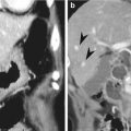
Fig. 23.4
Abdominal wall hematoma. (a) Transverse CT image shows expansion of right rectus sheath, within which high attenuation is identified, indicating blood clots (arrowheads). Extravasation (arrow) is also noted in the hematoma. (b) On selected angiogram of the right inferior epigastric artery, extravasation (arrow) is noted
23.5.5 Abdominal Wall Abscess in a Patient with Crohn’s Disease
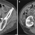
Fig. 23.5
Abdominal wall abscess in a patient with Crohn’s disease. Transverse CT image shows thickened segment of ileum (arrowheads) and diffuse intra-abdominal fat infiltration, suggesting inflammatory process. Abscess (asterisk) in the right lower quadrant is extending into the right abdominal wall, resulting in an abscess with air pocket (arrow)
23.5.6 Actinomycosis Involving Pelvic Cavity and Abdominal Wall
Fig. 23.6
Actinomycosis involving pelvic cavity and abdominal wall. (a) Transverse CT image shows a left adnexal mass (asterisk), which is extending to the left abdominal wall (arrowheads). Fat stranding around adjacent intra-abdominal fat suggests an inflammatory process. (b) Coronal CT image shows that the left adnexal mass (asterisk) is infiltrating to adjacent urinary bladder (arrowheads), which is markedly thickened. Due to these aggressive features, there may be difficulty in making the correct diagnosis on CT imaging
23.5.7 Desmoid Tumor in the Abdominal Wall
Fig. 23.7
Desmoid tumor in the abdominal wall. Transverse CT image shows a well-defined large mass (arrowheads) in the lower abdominal wall. The tumor shows heterogeneous attenuation. Urinary bladder is compressed by the tumor. Note the previous operation scar (curved arrows) by previous cesarean section