Abdominal Wall, Mesentery, Peritoneum, and Vessels


Abdominal Wall, Mesentery, Peritoneum, and Vessels


Michael S. Gee

Rahul A. Sheth

Salwa M. Haidar

Dilip Sankhla

Edward Y. Lee


INTRODUCTION

The abdominal wall, mesentery, peritoneum, and blood vessels are often overlooked anatomic regions on imaging studies, particularly in pediatric patients, in whom the focus of imaging studies is frequently the abdominal visceral organs. These regions have their own unique set of disorders with which radiologists should be familiarized. In addition, the spreading pattern of more common abdominal infectious, inflammatory, and neoplastic processes can include these areas, and their involvement may be the only imaging evidence of the underlying disease. This chapter reviews imaging techniques, relevant anatomy, and pathology pertaining to the abdominal wall, mesentery, peritoneum, and vessels in the pediatric population.


IMAGING TECHNIQUES


Radiography

The abdominal radiograph is often the best first-line imaging examination for the evaluation of pediatric abdominal diseases. Air within bowel loops provides intrinsic contrast that can be used to evaluate the caliber and distribution of bowel in diseases that lead to bowel obstruction. In addition, spaceoccupying lesions in the abdomen can displace bowel and appear on radiographs as regions of absent bowel gas. The radiograph is often an excellent “starting point” to exclude urgent or emergent complications, such as free intraperitoneal air. Although radiographs have poor soft tissue contrast resolution, they have high spatial resolution for detecting the presence of air and calcification within lesions. On a frontal radiograph, free intraperitoneal air is best seen with the patient positioned upright or in a decubitus position. A cross-table lateral radiograph can also be performed in sick infants who need to lie supine. Calcifications of the peritoneum, mesentery, or abdominal wall are also radiographically apparent. Abnormally displaced bowel loops or a localized paucity of bowel gas on an abdominal radiograph can be an indirect sign of ascites or a soft tissue mass in the peritoneum, retroperitoneum, or mesentery. A lateral abdominal radiograph can be helpful for detecting abdominal wall abnormalities, which may manifest as thickening, such as in cellulitis, or calcification. The abdominal vasculature is poorly evaluated on plain radiographs.


Ultrasound

Ultrasound (US) is often an initial imaging modality used in children because it does not require ionizing radiation, sedation, or anesthesia. US is helpful for evaluating children presenting with a palpable abdominal mass to identify the presence, location, and tissue composition of underlying lesions. In children, especially infants, the general lack of internal body fat facilitates imaging of abdominal lesions deep within the peritoneal cavity and retroperitoneum that might not be visible in adults because of acoustic attenuation or shadowing from adjacent bowel gas.

High-frequency (12 to 18 MHz) linear transducers are best for evaluating the subcutaneous tissues and abdominal wall, whereas lower-frequency (4 to 8 MHz) convex transducers are more suitable for evaluating the visceral organs, the peritoneal spaces, and small bowel mesentery.1 Harmonic imaging and compression technique can also be utilized to facilitate visualization of structures deep in the abdomen. Color and spectral Doppler US detect frequency shifts related to moving blood and are used to evaluate the arteries and veins for anatomic and flow-related abnormalities and to evaluate lesions seen on gray-scale US for internal vascularity to help discriminate complex cysts from solid masses.


Computed Tomography

Computed tomography (CT) is an imaging modality that is ideally suited for evaluating the abdominal wall, peritoneum, and mesentery, because of its high spatial resolution, cross-sectional imaging capability, and ability to provide highquality images of air-filled or calcified structures. Modern multirow detector CT scanners can image the entire abdomen of a child in ≤1 to 5 seconds and are well suited for imaging awake infants and toddlers who cannot suspend respiration or follow commands. In addition, CT scanners are available in most emergency rooms to evaluate acutely ill children at any time of day. However, an important concern with CT is the use of ionizing radiation, especially in pediatric patients. Prior to CT scanning for each child, an informed analysis that balances the diagnostic benefit of the examination against the potential risk of CT ionizing radiation should be undertaken.

Pediatric CT protocols of the abdomen and pelvis involve modification of a number of scan parameters that reduce radiation dose while still ensuring diagnostic image quality.2,3 These include setting limits on tube beam current (mA) and potential (kVp) depending on the study indication, increasing scan pitch, and utilizing thick sections (e.g., 2.5 to 5 mm) with retrospective thin collimation for multiplanar reformations. Modern CT scanners utilize adaptive tube current and voltage modulation to decrease radiation dose as a function of patient size. Routine abdominal CT scans are performed with intravenous contrast in the portal venous phase to optimize evaluation of the visceral organs. CT angiography is performed by acquiring images in the arterial phase of enhancement using either contrast bolus tracking or timing. Sagittal reformatted images are especially helpful for evaluating the subcutaneous tissues and abdominal wall as well as the ostia of the abdominal celiac and superior mesenteric arteries.


Magnetic Resonance Imaging

Magnetic resonance imaging (MRI) is being increasingly performed for pediatric abdominal evaluation. Its main advantages include the ability to acquire images in any plane, lack of ionizing radiation, and excellent soft tissue contrast resolution. Because of the lack of ionizing radiation, imaging of the same anatomic region may be performed at multiple time points following intravenous contrast material administration. This is helpful for evaluating blood vessels during combined MR angiography and venography as well as for characterizing the enhancement properties of masses. The superior soft tissue contrast resolution of MRI is ideally suited for tissue characterization of abdominal lesions detected on other imaging modalities, with T1-weighted MR images sensitive for the detection of fatty or hemorrhagic elements and T2-weighted MR images sensitive for cystic or soft tissue components.

Typical pediatric abdominal MRI protocols may include coronal single-shot fast spin-echo and balanced steady-state free precession MR images to provide a motion-free overview of abdominal anatomy as well as axial T1-weighted and fat-suppressed T2-weighted MR images to evaluate for pathology. 3D T1-weighted gradient recalled echo fat-saturated MR images are then acquired before and at multiple time points after intravenous gadolinium chelate administration to evaluate the abdominal vasculature and assess for enhancing lesions. For dedicated evaluation of the abdominal vasculature, time-resolved contrast-enhanced MR angiography/venography can be performed using dynamic acquisition of multiple 3D T1-weighted fast gradient recalled MR images to provide multiphase vascular imaging without the need to suspend respiration.4 The main disadvantages of MRI are its long scanning time (30 to 60 minutes for a typical examination) and motion-related degradation of image quality, which are both relevant to the pediatric population. Abdominal MRI in young children often requires conscious sedation or general anesthesia for these reasons. Nonetheless, MRI is commonly the study of choice for evaluating most abdominal wall, peritoneal, and vascular lesions prior to biopsy or surgical treatment.


Nuclear Medicine

Nuclear scintigraphy offers the advantage of radiotracer molecule specificity that can be used to characterize lesions based on physiologic properties. For example, 18F-flourodeoxyglucose (FDG) accumulates in cells with increased glucose metabolism and is often used in conjunction with positron emission tomography (PET) to determine malignancy/benignity of soft tissue lesions in the abdominal wall, mesentery, and peritoneum that are detected on other imaging modalities as well as imaging staging of many malignancies. 131I-MIBG is an adrenergic analog that accumulates in neuroblastoma cells and is used for primary neuroblastoma detection and staging. Nuclear scintigraphy using gallium (67Ga)-labeled or 111In-labeled white blood cells can be helpful for abscess detection in pediatric patients with suspected abdominal infection. 99mTc-labeled red blood cell scanning can help identify a mesenteric arterial bleeding source in pediatric patients with lower gastrointestinal bleeding.

Historically, the major limitations of nuclear scintigraphy have been poor spatial resolution and inability to anatomically localize detected abnormalities. However, the development of tomographic techniques, such as single photon emission computed tomography (SPECT) and PET, has been helpful in this regard, as well as the use of hybrid imaging of SPECT and PET with CT and MRI. Nuclear scintigraphy also involves long scan times similar to MRI and often requires sedation or general anesthesia to be performed in young children.


Conventional Angiography

Although most vascular pathology can be accurately diagnosed with noninvasive vascular imaging, there remains an important role for conventional angiography in the pediatric population. Continuous advancements in interventional techniques have expanded the diagnostic and therapeutic potential for angiography. Increasingly, complex percutaneous arterial interventions can be performed in children for a broad range of conditions including renovascular hypertension, transplant liver hepatic artery stenosis, and abdominal trauma.5

Awareness of radiation exposure to the pediatric patient and a commitment to the “ALARA” (“as low as reasonably achievable”)
principle are vitally important for the pediatric angiographer. Angiography, particularly during lengthy percutaneous interventions, has the potential to deliver the highest radiation dose of any imaging study. Methods to reduce radiation exposure during fluoroscopy include the use of pulse fluoroscopy, last-image hold, copper filtration, optimal collimating, and removal of anti-scatter grids while imaging neonates and small infants.

The most commonly accessed vessel in pediatric angiography is the common femoral artery.6 Arterial access in children can be challenging because of the relatively smaller size of blood vessels in this population; as such, vessel occlusion following catheterization is a more common occurrence in children than in adults. The umbilical artery remains patent for up to 5 days and can serve as suitable arterial access point. Young patients are particularly susceptible to volume overload and nephrotoxicity from iodinated contrast media, and so, the volumes of fluids and contrast media used should be closely monitored.


NORMAL ANATOMY


Abdominal Wall

The anterior abdominal wall extends cranially to the xiphoid process, laterally to the rib cage, and caudally to the iliac and pubic bones. The anterior abdominal wall contributes to respiration as well as urination, defecation, and coughing. The muscles of the anterior abdominal wall also assist with flexion and extension of the body at the hips. These muscles include the rectus abdominis anteriorly and the external oblique, internal oblique, and transversus abdominis laterally/posterolaterally.


FIGURE 20.1 Diagram of peritoneal spaces.


Peritoneum, Peritoneal Spaces, and Mesentery

The peritoneum is the largest serous membrane in the body. It is a thin, translucent single sheet of mesothelial tissue. Microscopically, the peritoneum is composed of flat mesothelial cells. The peritoneum that invests abdominal organs is termed the visceral peritoneum, and the peritoneum that lines the abdominal cavity is known as the parietal peritoneum. The potential space between these two layers of the peritoneum is normally filled with a small volume of serous fluid, minimizing friction when the two layers contact. In boys, the peritoneal cavity is a closed space. In girls, however, the peritoneal cavity is pierced laterally by the fallopian tubes.

The peritoneal cavity is segregated into discrete compartments by peritoneal ligaments, structures that represent double layers of the visceral peritoneum and provide suspensory support for abdominal organs (Figs. 20.1 and 20.2). The two major compartments of the peritoneal cavity as defined by
the peritoneal ligaments are the greater sac and the lesser sac, also known as the omental bursa.


FIGURE 20.2 A 13-year-old girl on peritoneal dialysis undergoing CT peritoneogram to assess for peritoneal adhesions. Contrast opacification of the peritoneum identifies the major spaces and ligaments. RSP, right subphrenic space; LSP, left subphrenic space; GHL, gastrohepatic ligament; HDL, hepatoduodenal ligament; LS, lesser sac; S, stomach; P, pancreas; D, duodenum; RPC, right paracolic space; LPC, left paracolic space; PS, pelvic space. (Image courtesy of Jonathan R. Dillman, MD, MSc, Cincinnati Children’s Hospital Medical Center, Cincinnati, OH.)

Two of the most important peritoneal ligaments are the omentum and the mesentery. The omentum is subdivided into the greater and lesser omentum, the latter of which is composed of the gastrohepatic and hepatoduodenal ligaments. The lesser omentum fixes the stomach and first portion of the duodenum to the liver. The gastrohepatic ligament binds the lesser curvature of the stomach to the liver, containing the coronary vein and left gastric artery. The hepatoduodenal ligament connects the duodenal bulb to the liver and contains the portal vein, hepatic artery, common hepatic bile duct, and a portion of the cystic duct. The greater omentum, also known as the gastrocolic ligament, hangs dependently from the greater curvature of the stomach, anterior to the small bowel.

The mesentery is a double layer of peritoneum that ensconces abdominal viscera and attaches them to a fixed anatomic structure, typically the abdominal wall. Although the mesentery is a complex three-dimensional structure that undergoes extensive rotation, translation, and resorption during development, it is entirely composed of an uninterrupted sheet of tissue. Nonetheless, by convention, the mesentery is divided into several discrete mesenteries. The small bowel mesentery runs from the ligament of Treitz in the left upper quadrant to the ileocecal valve in the right lower quadrant. It binds the small bowel to the retroperitoneum and contains the superior mesenteric arteries and veins. The transverse mesocolon is a broad fold of peritoneal lining that binds the transverse colon with the posterior abdominal wall. It is continuous with the posterior layers of the greater omentum and contains the middle colic vessels. Similarly, the sigmoid mesocolon binds the sigmoid colon to the posterior pelvic wall, containing the hemorrhoidal and sigmoid arteries and veins.

There are multiple additional peritoneal ligaments. These include the falciform ligament, which represents the vestigial remnant of the embryologic ventral mesentery. It contains the obliterated umbilical vein and separates the subphrenic space into left and right compartments. The gastrosplenic ligament arises from the embryologic dorsal mesentery and connects the greater curvature of the stomach with the spleen. The splenorenal ligament also arises from the embryologic dorsal mesentery and can contain splenorenal shunt vasculature in the setting of portal hypertension.7

The omenta, mesenteries, and other ligaments segregate the peritoneal cavity into multiple, somewhat discrete spaces, serving as both barriers and pathways for the spread of disease (Table 20.1). Knowledge of these spaces is indispensable for understanding common patterns of spread of infectious, inflammatory, or neoplastic processes in the abdomen. It should be noted, however, that while the peritoneal ligaments do serve as boundaries for these spaces, the boundaries are not absolute and they can be overcome.

The transverse mesocolon separates the peritoneal cavity into the bilateral supramesocolic and inframesocolic spaces. Laterally, the peritoneal cavity contains the paired paracolic gutters. Inferiorly, the peritoneal cavity is composed of the pelvic space.


TABLE 20.1 Anatomic Spaces of the Peritoneal Cavity


Supramesocolic


Left supramesocolic


Left perihepatic


Left subphrenic


Perisplenic


Right supramesocolic


Right subphrenic


Subhepatic (Morison’s pouch)


Lesser sac


Inframesocolic


Left inframesocolic


Right inframesocolic


Paracolic


Left paracolic


Right paracolic


Pelvic


Reference: Tirkes T, Sandrasegaran K, Patel AA, et al. Peritoneal and retroperitoneal anatomy and its relevance for cross-sectional imaging. Radiographics. 2012;32(2):437-451.


The left supramesocolic space is somewhat arbitrarily subdivided into the perihepatic, left subphrenic, and perisplenic spaces. The right supramesocolic space contains the right subphrenic and subhepatic spaces (also known as Morison pouch) and the lesser sac. The bilateral supramesocolic spaces typically communicate freely, with the falciform ligament often representing an incomplete barrier between the left and right subphrenic spaces. The left supramesocolic space also typically communicates with the left paracolic gutter, with the phrenicocolic ligament serving as an incomplete barrier; the right paracolic gutter is a continuation of the right subhepatic space. Both paracolic gutters extend into the pelvic space.

The bilateral inframesocolic spaces, on the other hand, do not communicate with the paracolic gutters because of the right and left colon. The left inframesocolic space is in communication with the pelvic space; the right inframesocolic space, on the other hand, is small and isolated from the pelvis by the small bowel mesentery.

Characterizing lesions as intraperitoneal or extraperitoneal is often highly relevant to the surgeon but can be a challenge for the radiologist. Adding to the challenge is the convention of using “intramesenteric” and “intraperitoneal” interchangeably. Strictly speaking, the intraperitoneal space refers to a thin potential space between the layers of parietal and visceral pleura; the intramesenteric space, on the other hand, describes blood vessels, lymph nodes, and fat that are encased by but not contained within visceral peritoneum. The intramesenteric space is in fact in continuity with the extraperitoneal space including the retroperitoneum. For this reason, the term “subperitoneal space,” which includes the intramesenteric space and extraperitoneal space, may be a more helpful term to the surgeon, as accessing lesions in this space does not require violation of the peritoneum.8


Abdominal Vessels


Abdominal Aorta

The abdominal aorta extends from the diaphragmatic hiatus at approximately the T12-L1 interspace to the pelvis where it bifurcates into the bilateral common iliac arteries at approximately the L4 level (Fig. 20.3). The diameter of the aorta narrows as it descends in the abdomen and provides mesenteric and visceral branches. The development of the aorta begins in the 3rd week of embryogenesis. Many dorsal and ventral segmental arteries arise from the primitive aorta, some of which regress during development, and others of which persist.9


Inferior Vena Cava

The formation of the inferior vena cava (IVC) (Fig. 20.4) and its major tributaries is a complex process, involving three paired venous systems that selectively regress and fuse. Three retroperitoneal venous systems form chronologically during weeks 6 through 8 of embryogenesis. The posterior cardinal system is the earliest such system, arising in week 6, and does not contribute to the normal IVC. The subcardinal system forms in week 7 and provides the prerenal segment of the IVC. The supracardinal system is the last system to form, arising during week 8, and contributes the postrenal segment of the IVC. Anastomosis between the subcardinal and supracardinal systems form the renal segment of the IVC.10 An appreciation of this complex embryologic development provides a rational foundation for the array of stereotyped anomalies described subsequently in this chapter.


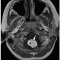
FIGURE 20.3 Normal angiographic anatomy of the abdominal aorta and its visceral branches. Arrows indicate the major abdominal aortic visceral branches. CHA, common hepatic artery; SA, splenic artery; RRA, right renal artery; LRA, left renal artery; SMA, superior mesenteric artery.


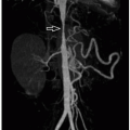
FIGURE 20.4 Normal inferior vena cavogram. The ostia of the renal veins are identified as jets of unopacified blood (arrows) flowing into the inferior vena cava.


Mesenteric Arteries

The mesenteric arteries develop during embryogenesis from primitive ventral segmental arteries. All but three of these arteries are resorbed and lead to the celiac axis, superior mesenteric artery (SMA), and inferior mesenteric artery (IMA). The celiac axis supplies the foregut and arises from the 10th segmental artery; the SMA supplies the midgut and arises from the 13th segmental artery; the IMA supplies the hindgut and arises from either the 21st or 22nd segmental artery.9 The majority of vascular anatomic variants involving the mesenteric arteries reflect incomplete resorption of the primitive arteries.

The SMA (Fig. 20.5) arises ˜1 cm below the origin of the celiac axis, typically at the level of the L1 vertebral body. It provides blood flow to the duodenum, jejunum, ileum, right colon, and majority of the transverse colon. Several structures normally course between the SMA and the abdominal aorta, including the third portion of the duodenum and the left renal vein. Physical compression of these structures can lead to SMA syndrome and nutcracker syndrome, respectively.

One of the first right-sided branches off the SMA is the inferior pancreaticoduodenal artery, which anastomoses with the superior pancreaticoduodenal artery arising from the gastroduodenal artery, thus providing an important celiac axis-SMA collateral pathway (Table 20.2). This pancreaticoduodenal arcade nourishes the pancreatic head as well as the duodenum. The inferior pancreaticoduodenal artery more commonly arises as two separate branches, namely, the antero- and posteroinferior pancreaticoduodenal arteries, but it can also branch off as a single trunk that subsequently bifurcates.11


FIGURE 20.5 Normal angiographic anatomy of the superior mesenteric artery (SMA) and its branches. On the right, the inferior pancreaticoduodenal artery (IPDA) anastomoses with the superior pancreaticoduodenal artery branch of the gastroduodenal artery, forming a collateral pathway between the SMA and celiac axis. There are multiple left-sided jejunal branches (JB). On the right are the middle colic (MCA), right colic (RCA), and ileocolic arteries (ICA), and anastomoses among these arteries form a marginal artery (of Drummond) that irrigates the right and transverse colon. Distal to the origin of the ICA, multiple ileal branches (IB) supply the ileum.


TABLE 20.2 Collateral Pathways of Mesenteric Arteries


Celiac axis to SMA


  1. 1. Arc of Buehler (persistent embryonic communication between celiac axis and SMA)


  2. 2. Superior pancreaticoduodenal artery to inferior pancreaticoduodenal artery


SMA to IMA


  1. 1. Marginal artery of Drummond (anastomotic connections between middle colic and left colic arteries along the mesenteric margin of the large bowel)


  2. 2. Arc of Riolan or meandering mesenteric artery (short direct anastomosis between middle colic and left colic arteries)


IMA to internal iliac artery


  1. 1. Superior rectal (hemorrhoidal) to middle and inferior rectal (hemorrhoidal) arteries


SMA, superior mesenteric artery; IMA, inferior mesenteric artery.


Following the inferior pancreaticoduodenal artery, the next right-sided branch is the middle colic artery, followed by the right colic artery and ileocolic artery. The ileocolic artery serves as the landmark beyond which all subsequent small branches off the SMA supply the ileum and no longer the jejunum. The SMA provides ˜4 to 6 left-sided jejunal branches and 9 to 13 ileal branches. The middle colic artery nourishes the transverse colon and provides collateral circulation via anastomotic communications with the IMA. It gives rise to a right branch, which anastomoses with the right colic artery, and a left branch, which in some patients anastomoses with the left colic artery (via the arc of Riolan or meandering mesenteric artery) along the root of the small bowel mesentery and as an important SMA-IMA collateral pathway. The middle colic artery typically arises from the SMA before the artery pierces the mesentery. The origin of the middle colic artery, however, can be variable, having been identified as arising from the celiac axis, common hepatic artery, and splenic artery; it may also arise from a replaced right hepatic artery or the gastroduodenal artery. Branches of the middle, right, and ileocolic arteries form a marginal artery (also known as the marginal artery of Drummond) along the inner border of the colon that nourishes the ascending colon and connects with branches of the IMA. Importantly, however, the right colic artery is commonly absent. The middle and right colic artery may share a single trunk off of the SMA.

The IMA (Fig. 20.6) arises from the ventral abdominal aorta below the origin of the SMA at the level of L3.
This artery is the smallest of the mesenteric arteries and nourishes the distal transverse colon, descending colon, sigmoid colon, and rectum. The major branches of the IMA are the left colic, sigmoid, and hemorrhoidal arteries, all of which branch off to the left. The left colic ascends from its origin off the IMA to anastomoses with branches from the SMA. In ˜12% of individuals, the left colic artery is absent. In this situation, the perfusion of the descending and sigmoid colon is provided by the colosigmoid artery. Occasionally, the left colic artery may arise from the SMA. The left colic artery extends cephalad to the splenic flexure in the majority of patients and reaches the mid-aspect of the transverse colon in ˜15% to 20% of patients. However, perfusion of the splenic flexure is highly variable, and in some patients, the middle colic artery may be the only artery irrigating this territory.


FIGURE 20.6 Normal angiographic anatomy of the inferior mesenteric artery (IMA). The left colic artery (LCA) anastomoses with the middle colic artery of the superior mesenteric artery (SMA) to provide SMA-IMA collaterals. Multiple sigmoid branches (SB) arise from the IMA, and the terminal branch of which is the superior hemorrhoidal artery (SHA).


Mesenteric Veins

The superior mesenteric vein (SMV) (Fig. 20.7) receives blood from multiple mesenteric veins, including the ileocolic, gastrocolic, right colic, and middle colic veins. These veins typically merge into a single trunk that joins with the splenic vein to form the portal vein. Occasionally, however, the tributaries may not coalesce into a single trunk but rather into right and left mesenteric branches, which then join the splenic vein.11,12

The inferior mesenteric vein (IMV) receives blood from the superior hemorrhoidal vein, sigmoid vein, and left colic vein. The IMV may drain into the SMV or continue cephalad to drain into the splenic vein or splenoportal confluence.11,12


FIGURE 20.7 Superior mesenteric vein (SMV) appearance on CT. Maximum intensity projection reconstruction CT image demonstrates the normal anatomy of the SMV. This vein receives blood from multiple mesenteric veins, which coalesce most commonly into a single trunk that subsequently joins with the splenic vein to form the portal vein (PV).


SPECTRUM OF ABDOMINAL WALL DISORDERS


Congenital and Developmental Anomalies


Omphalocele and Gastroschisis

Omphalocele refers to herniation of the abdominal viscera through the umbilical cord. The most common organs within an omphalocele are the liver and small bowel, with the spleen, stomach, colon, and bladder being less common.13 It arises from a defect caused by failure of central migration of the lateral mesodermal folds destined to become the anterior abdominal wall. The lining of an omphalocele is composed of peritoneum and amnion with interposed Wharton jelly. Although the majority of omphaloceles are isolated, there is high association with chromosomal abnormalities. Omphaloceles are classified based on size, with size <5 cm associated with higher survival rate (>80%) and having a higher association with chromosomal abnormalities and isolated small bowel herniation, whereas size ≥5 cm is associated with lower survival rate (<50%) and a higher likelihood of having liver herniation and pulmonary hypoplasia.

On physical examination, the umbilical cord is seen at birth to insert into the mass. Plain radiographs show an anterior mass extending from the midline abdominal wall, with herniated bowel loops showing gas within the hernia.
Typically an omphalocele is diagnosed on prenatal US showing an anterior midline herniation with a covering membrane (Fig. 20.8) and the umbilical cord vessels seen inserting at the base of the hernia.


FIGURE 20.8 Imaging features of omphalocele and gastroschisis. A: Abdominal radiograph of a neonatal boy with omphalocele demonstrates a large anterior abdominal wall hernia (arrow) containing the stomach (asterisk) and multiple bowel loops. B: Prenatal sagittal T2-weighted MRI of a fetal omphalocele at 32 weeks of gestational age demonstrates an anterior abdominal hernia containing the liver covered by a thin outer membrane (asterisk), with the umbilical cord vessels inserting at the hernia base (arrow). In contrast, prenatal imaging of a 25-week-gestational-age fetus with gastroschisis on ultrasound (C) and sagittal T2-weighted MR image (D) demonstrate anterior herniation of small bowel loops (arrowheads) with no outer covering membrane. (Radiograph [A] provided by Jonathan R. Dillman, MD, MSc, Cincinnati Children’s Hospital Medical Center, Cincinnati, OH. Ultrasound [C] and MRI [B and D] images provided by Sudha Anupindi, MD, and Teresa Victoria, MD, Children’s Hospital of Philadelphia, Philadelphia, PA.)

Gastroschisis is a congenital defect in the anterior wall that is off-midline in the paraumbilical location, typically to the right of midline. In contrast to omphalocele, there is a full-thickness defect in the abdominal wall, with herniated abdominal contents contained only in amniotic fluid without a covering membrane. Gastroschisis can produce bowel injury depending on the amount and duration of bowel herniation. Herniated intestine can become edematous and ischemic because of exposure to amniotic fluid, and blood supply can be impaired by the neck of the abdominal wall defect. Gastroschisis is associated with intestinal atresia (usually jejunal or ileal) and gastrointestinal motility disorders. Closed gastroschisis, or “vanishing midgut,” refers to midgut infarction resulting from abdominal wall defect closure around herniated bowel.14

Prenatal US and MRI show herniated intestine with no covering membrane (Fig. 20.8C, D) and a normal umbilical cord vessel insertion distinct from the site of herniation. Elevated alpha-fetoprotein (AFP) level is present in maternal serum in over 90% of cases.

The current management of omphalocele and gastroschisis is surgical repair, which consists of placing the herniated organs back into the abdomen cavity (often covered by a protective silo) followed by complete closure of the defect via either one operation or gradual staged operations.


Prune Belly Syndrome

Prune belly (Eagle-Barrett) syndrome is characterized by a defect in abdominal wall musculature, urinary system ectasia, and cryptorchidism. It is a rare disorder with a male predominance and estimated incidence of 3 to 4 per 100,000.15 The etiology is thought to be in utero urethral obstruction due to either a hypoplastic prostate or a failure in
mesodermal development, leading to urinary tract distention, abnormal abdominal wall muscular development, and failure of testicular descent.16,17 The abdominal wall defect classically consists of disorganized central abdominal wall muscles that are infiltrated with collagen bundles.18 Cryptorchidism has been attributed to both impeded testicular descent by the distended urinary system as well as atresia of the gubernaculum, which does not pull adequately.16


FIGURE 20.9 Prune belly syndrome in a newborn boy. A: Radiograph demonstrates marked abdominal wall laxity. B and C: Renal ultrasound images show bilateral hydronephrosis. The kidneys are echogenic and contain several small cysts, suggestive of underlying renal dysplasia.

Diagnosis of prune belly syndrome is typically made during the antenatal period, with fetal imaging demonstrating bilateral hydroureteronephrosis, bladder distention, and oligohydramnios. The appearance can be similar to posterior urethral valves, although typically lacking prostatic urethral dilation seen with PUV.16 Severe renal dysfunction associated with prune belly syndrome is associated with pulmonary hypoplasia and cardiovascular dysfunction, and perinatal mortality rates are in the 10% to 25% range depending on the degree of prematurity and cardiopulmonary compromise.16 Because of this, antenatal urinary decompression techniques such as vesicoamniotic shunting are considered in some severe cases in an attempt to improve perinatal survival.19 Postnatal imaging appearance includes severe abdominal distention and wall laxity on radiographs (Fig. 20.9A), as well as hydroureteronephrosis (Fig. 20.9B), renal dysplasia, and cryptorchidism on US.

Treatment of prune belly syndrome includes managing the lower urinary tract, abdominal wall, and cryptorchidism.16 The goal of urinary management is to preserve renal function and avoid urinary tract infection, via either conservative methods or surgical reconstruction. Premature renal failure is a common long-term complication, with approximately one-third of affected patients requiring renal transplant later in life.20 Abdominal wall surgical reconstruction aids with abdominal tone and can improve bladder voiding through augmented sensation and contraction.21 Bilateral orchidopexy is standardly performed as well.


Proteus Syndrome

Proteus syndrome is a tissue overgrowth disorder that is due to a mutation in the AKT1 gene leading to activation of the PI3K-AKT signaling pathway22 and overgrowth of multiple tissue types. Characteristic lesions include cerebriform connective tissue nevi, vascular malformations, focal gigantism of a digit or limb, facial dysmorphism, dysregulated adipose tissue (lipomas or lipohypoplasia), pulmonary bullae, and deep vein thrombosis.23 It is a sporadic syndrome that is characterized by mosaic distribution of lesions and progressive disproportionate tissue overgrowth, which in the limbs and digits consists predominantly of bony overgrowth with cortical thinning and relative paucity of overlying soft tissue.24

Proteus syndrome is a rare disorder only affecting several hundred patients in the United States and Western Europe, with a male predominance and physical manifestations appearing either at birth or later in childhood.23,25 Because the mutation is poorly detected in peripheral blood, diagnosis is made predominantly by the presence of multiple disease defining lesions.

Imaging plays an important role in establishing the diagnosis in patients with suspected Proteus syndrome, including a radiographic skeletal survey as well as targeted MRI and/or CT of clinically affected areas or cross-sectional imaging of the chest, abdomen, and pelvis in asymptomatic patients.23,24 Common abdominal wall abnormalities include vascular malformations and fatty lesions (Fig. 20.10).

Pediatric patients with Proteus syndrome experience numerous disease complications over their lifetime, including large joint arthrosis and scoliosis, visceral organ overgrowth, and development of tumors including ovarian cystadenomas and parotid adenomas.25 Deep venous thrombosis and pulmonary embolism are other common disease complications.23 Imaging is helpful for detecting and characterizing these various disease manifestations. The differential diagnosis for Proteus syndrome includes: PTEN hamartoma tumor syndrome, CLOVE syndrome, and Klippel-Trenaunay syndrome.


Infectious and Inflammatory Disorders


Abdominal Wall Cellulitis and Abscess

Cellulitis is acute bacterial infection of the skin and subcutaneous tissues. It is associated with features of acute inflammation including erythema, swelling, warmth, and tenderness to palpation.26 Often the area of involvement spreads over time and may
be accompanied by fever or malaise. Most commonly, cellulitis is a complication of an overlying skin disorder, such as a penetrating wound (including a puncture, abrasion, or bite), ulcer (e.g., varicella lesions or newborn omphalitis), or dermatosis.27,28 Abdominal wall cellulitis in children may also be a sequela of abdominal surgery, such as appendectomy. Most cases of cellulitis are due to infection by Streptococcus and Staphylococcus skin flora. Less typical organisms include oral flora in cases of cellulitis secondary to a bite or Gram-negative rods, anaerobic flora, and fungi in immunocompromised patients.28


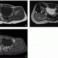
FIGURE 20.10 Proteus syndrome in an 15-year-old boy. Axial enhanced CT images demonstrate a vascular malformation (A; arrows) in the right flank abdominal wall as well as a large abdominal wall lipoma (B; asterisk).

The diagnosis of cellulitis is typically made based on cutaneous examination and clinical history, without the need to perform imaging or obtain a skin culture.28 Imaging can be performed in two specific clinical scenarios, namely, US to exclude an associated abscess requiring drainage and MRI to exclude necrotizing fasciitis, which would necessitate surgical debridement. CT may also be performed to evaluate for the presence of gas within the soft tissues that can suggest the diagnosis of necrotizing fasciitis. US features of cellulitis include edema and thickening of the skin and subcutaneous tissues, with an associated abscess appearing as a hypoechoic focal fluid collection with posterior acoustic enhancement and often an echogenic rim (Fig. 20.11). The presence of an abscess typically is an indication for surgical or image-guided drainage, as antibiotic therapy is typically inadequate to eradicate the abdominal wall abscess.


FIGURE 20.11 Abdominal wall cellulitis in a newborn boy. A: Gray-scale ultrasound image of the paraumbilical region demonstrates heterogeneous thickening of the paraumbilical abdominal wall soft tissues, consistent with cellulitis. B: Color Doppler ultrasound image shows mild hyperemia.

MRI is sometimes performed in cases of rapidly progressive skin infection to evaluate for necrotizing fasciitis, which is a surgical emergency. MRI features of necrotizing fasciitis include abnormal thickening, fluid, and enhancement of the deep fasciae.29 MRI appears to be sensitive but not specific for necrotizing fasciitis, and generally, it is not recommended if there is strong clinical suspicion.28,29 The typical treatment of uncomplicated abdominal wall cellulitis is antibiotic therapy.


Neoplastic Disorders


Benign Neoplasms


Desmoid Tumor

Desmoid tumors, also known as deep or aggressive fibromatosis, are fibrous mesenchymal neoplasms that are locally aggressive but do not exhibit distant metastasis.30 These rare tumors
(<5 per million annual incidence) arise mostly in young adults and less commonly in children, with a slight female predominance and peak incidence in the third and fourth decades.31 Desmoid tumor locations are traditionally classified as intraabdominal (mesenteric or pelvic), abdominal wall, or extra-abdominal (most commonly in the proximal extremities, head, and neck). Patients with Gardner-type familial adenomatous polyposis (FAP) have a >800-fold increased incidence of desmoids, which are usually intra-abdominal or mesenteric in location.32,33 Abdominal wall desmoids occurring in the rectus abdominis or internal oblique muscles have an association with pregnancy.34 Desmoid tumors usually occur as single tumors, although ˜15% are multiple.34


FIGURE 20.12 Abdominal wall desmoid tumor in a 17-year-old boy with Gardner syndrome. Sagittal T2-weighted (A) and postcontrast T1-weighted fat-saturated (B) MR images demonstrate a hyperintense and enhancing mass (arrows) centered in the rectus abdominis muscle, consistent with known desmoid tumor.

Desmoid tumors, especially those in the abdominal wall or extra-abdominal locations in young patients, usually present as a slowly growing palpable mass and often are evaluated initially by US. The sonographic appearance of a desmoid is a well-defined hypoechoic mass with variable internal Doppler vascularity.35 MRI is frequently performed as the next imaging modality in young patients to define lesion extent and relationship to adjacent structures (Fig. 20.12). Desmoid tumors demonstrate variable T2-weighted signal intensity depending on the degree of collagen and myxoid matrix deposition; collagen-rich tumors are associated with signal hypointensity, and myxoid matrix-rich tumors are associated with signal hyperintensity.30,36 The lesions demonstrate variable enhancement with intravenous contrast material administration for the same reason.

Grossly, desmoid tumors are firm and usually fairly well circumscribed. They tend to have a white-gray whorled cut surface (Fig. 20.13). Microscopically, fascicles of fibroblasts in a variably collagenized matrix are seen. Genetically, the tumors often show trisomy of chromosome 8; mutations in the beta-catenin or APC gene may also be seen.37

Abdominal wall desmoids are often amenable to percutaneous needle biopsy because of their superficial location. Patients diagnosed with desmoid tumor should be screened for FAP, as 2% of all desmoid tumors are FAP associated.32 Asymptomatic desmoid tumors that are stable over time may be observed, although for abdominal wall desmoids, surgical resection with wide margins with or without abdominal wall reconstruction is the preferred approach and is associated with a very low local recurrence rate.31,32,38,39 Systemic therapy or radiation can also be considered for cases deemed nonresectable.31,32


Malignant Neoplasms


Soft Tissue Sarcoma

Soft tissue sarcomas arising within the abdominal wall are rare and account for only 1% to 5% of total soft tissue sarcomas.39 Soft tissue sarcomas as a whole are uncommon in the pediatric population, with <1,000 cases in the United States each year.40 Sarcomas constitute the most common primary abdominal wall malignancy in patients of all ages.41
In children, soft tissue sarcomas occurring in the abdominal wall are primarily nonrhabdomyosarcoma soft tissue sarcomas (NRSTS). Such NRSTS are a heterogeneous group of histologic subtypes derived from mesenchymal cells, which often have characteristic genetic translocations that aid in their diagnosis.40 This includes infantile fibrosarcoma, which is a rare NRSTS that occurs specifically in infants under 2 years of age (median age 3 months) and presents typically as a large infiltrative soft tissue mass with associated skin discoloration that may mimic vascular malformation.42 Synovial sarcoma (Fig. 20.14) is one of the common sarcomas in older children. All NRSTS typically present as a slowing growing and painless mass.


FIGURE 20.13 Gross appearance of desmoid fibromatosis developing after ileostomy closure in an 18-year-old man with familial adenomatous polyposis. The 5 cm mass ( asterisk) is firm and pale tan, distinct from the red-brown abdominal musculature.


FIGURE 20.14 Gross appearance of a synovial sarcoma from the abdominal wall of a 13-year-old boy. This is a 6.5 cm tumor resected after chemotherapy. The tumor (asterisk) is multilobular and light tan with soft pale areas of necrosis.

The initial evaluation of abdominal wall soft tissue sarcoma is similar to the abdominal wall desmoid management, including US43 to confirm the presence of a solid soft tissue mass and identify its location, followed by CT or MRI to assess extent of invasion and relationship to nearby neurovascular structures, aiding in surgical excision planning (Fig. 20.15).44 Percutaneous needle or surgical incisional biopsy is the mainstay of histologic diagnosis of soft tissue sarcomas. Once the diagnosis is made, staging CT of the chest, abdomen, and pelvis (with or without 18F-FDG-PET) is routinely performed to assess for metastatic disease.45 Surgical excision is the mainstay of abdominal wall sarcoma treatment but is often combined with radiation therapy and/or chemotherapy,44 with potential toxicities to abdominal organs balanced against lower likelihood of local recurrence.41


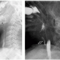
FIGURE 20.15 Abdominal soft tissue sarcoma in a 17-year-old boy. A: Sagittal enhanced CT image demonstrates a soft tissue mass (arrow) in the abdominal wall. B: Ultrasound depicts the lesion as a hypoechoic mass (calipers) with internal Doppler vascularity. C: Ultrasound-guided percutaneous core needle biopsy of the lesion established the diagnosis of synovial sarcoma.


Metastasis

Abdominal wall metastasis in the pediatric population is very rare. Pediatric malignancies that have been shown to metastasize to the skin or subcutaneous soft tissues include neuroblastoma, Wilms tumor, rhabdomyosarcoma, synovial sarcoma, and angiosarcoma46,47 (Fig. 20.16). Diffuse subcutaneous metastases in neuroblastoma account for the so-called blueberry muffin appearance. Although most cases of abdominal wall metastasis are attributable to hematogenous tumor spread (Figs. 20.16 and 20.17), a few cases of abdominal wall “metastasis” in adults have also been attributable to implantation after surgical resection of an intra-abdominal malignancy.41 The diagnosis of metastasis should be considered in any pediatric patient with a new abdominal wall mass and a history of prior malignancy.


Traumatic Disorders


Traumatic Abdominal Wall Injuries and Hernias

Traumatic abdominal wall hernia (TAWH) is the disruption of abdominal wall muscle and fascia in the absence of skin penetration, secondary to blunt trauma. This occurs in ˜1% of all blunt trauma and up to 9% of blunt trauma patients undergoing abdominal CT.48 In children, these injuries occur
primarily as a result of high-energy lap-belt trauma during motor vehicle collisions or from low-energy falls onto bicycle handlebars (the so-called handlebar hernia).49,50,51 The vast majority of these injuries that are bicycle related are in boys, with a mean age of ˜10 years.51

Only gold members can continue reading. Log In or Register to continue

Stay updated, free articles. Join our Telegram channel

Oct 13, 2018 | Posted by in PEDIATRIC IMAGING | Comments Off on Abdominal Wall, Mesentery, Peritoneum, and Vessels

Full access? Get Clinical Tree

Get Clinical Tree app for offline access