Abnormalities of the Deltoid Ligament
Anatomic Considerations
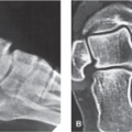
The ankle is a hinge-type articulation between the distal tibia, the two malleoli, and the talus (Fig. 5.1). The articular surface is covered with hyaline cartilage, which is susceptible to arthritis. The joint is surrounded by a dense capsule that helps strengthen the ankle. The joint capsule is lined with a synovial membrane that attaches to the articular cartilage. The ankle joint is innervated by the deep peroneal and tibial nerves.
The major ligaments of the ankle joint include the deltoid, anterior talofibular, calcaneofibular, and posterior talofibular ligaments, which provide the majority of strength to the ankle joint (Fig. 5.2). The deltoid ligament is exceptionally strong and is not so subject to strain as the anterior talofibular ligament. The triangular-shaped deltoid ligament is made up of a number of smaller separate ligaments including the anterior tibiotalar ligament, tibiocalcaneal ligament, posterior tibiotalar ligament, and tibionavicular ligament (Fig. 5.3). These ligaments are arranged in two layers. Both layers attach above to the medial malleolus. A deep layer attaches below to the medial body of the talus, with the superficial fibers attaching to the medial talus, the sustentaculum tali of the calcaneus, and the navicular tuberosity (Fig. 5.4).
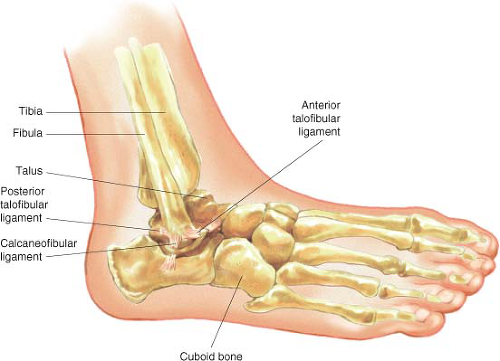 FIGURE 5.1 The articulations of the ankle joint. |
Clinical Correlates
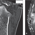
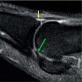
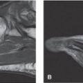
One of the four major ligaments of the ankle joint, the deltoid ligament is a strong, triangular-shaped, bilaminar ligament that runs from the medial malleolus, with the deep layer of the ligament attaching below to the medial body of the talus, and the superficial layer of the ligament attaching to the medial talus, the sustentaculum tali of the calcaneus, and the navicular tuberosity (Fig. 5.3). Also known as the medial ligament of talocrural joint, the deltoid ligament is susceptible to strain at the joint line or avulsion at its origin or insertion. The deltoid ligament is frequently injured from eversion injuries to the ankle that occur when tripping when wearing high heels, landing hard on uneven surfaces, and during dancing, soccer, and American football (Fig. 5.4). The pain of deltoid ligament damage is localized to the medial ankle and is made worse with plantar flexion and eversion of the ankle joint. Significant swelling and ecchymosis
are often evident after acute injury (Fig. 5.5). Activity, especially involving weight bearing, plantar flexion, and eversion of the ankle will exacerbate the pain. Local heat and decreased activity as well as elevation of the affected ankle may provide a modicum of relief. Sleep disturbance is common in patients suffering from trauma to the deltoid ligament of the ankle. Coexistent fracture, bursitis, tendinitis, arthritis, or internal derangement of the ankle may confuse the clinical picture after trauma to the knee joint making clinical diagnosis difficult.
are often evident after acute injury (Fig. 5.5). Activity, especially involving weight bearing, plantar flexion, and eversion of the ankle will exacerbate the pain. Local heat and decreased activity as well as elevation of the affected ankle may provide a modicum of relief. Sleep disturbance is common in patients suffering from trauma to the deltoid ligament of the ankle. Coexistent fracture, bursitis, tendinitis, arthritis, or internal derangement of the ankle may confuse the clinical picture after trauma to the knee joint making clinical diagnosis difficult.
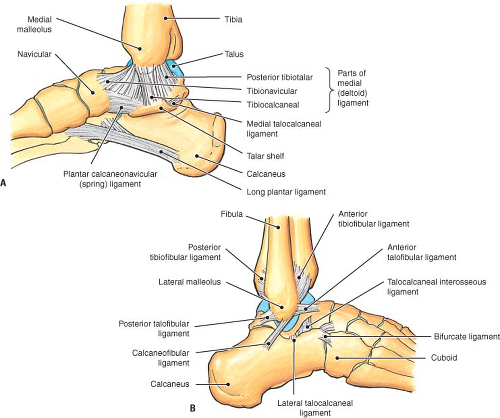 FIGURE 5.2 A, B: The anatomy of the deltoid ligament and its relationship with the other ligaments of the medial ankle. |
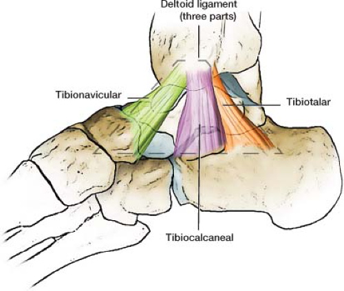 FIGURE 5.3 The triangular-shaped deltoid ligament is made up of a number of smaller separate ligaments, including the anterior tibiotalar ligament, tibiocalcaneal ligament, posterior tibiotalar ligament, and tibionavicular ligament. |
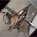
 FIGURE 5.4 Medial view of one osteoarticular dissection showing the components of the superficial layer of the deltoid ligament, especially the tibiospring ligament (major component). (1) Tibiospring ligament. (2) Spring ligament (superomedial calcaneonavicular ligament). (3) Posterior tibial tendon (cut). (4) Medial malleolus. (5) Sustentaculum tali. (6) Navicular tuberosity. (7) Lateral process of the talus. (8) Medial process of the talus. (9) Tibionavicular ligament (major component). (10) Superficial tibiotalar ligament or superficial posterior tibiotalar ligament (additional band). (11) Medial cuneonavicular ligament. (12) Long plantar ligament. (13) Plantar calcaneocuboid ligament. (14) Medial talocalcaneal ligament. (15) Medial cuneiform. (16) Calcaneal or Achilles tendon (cut). (From Hintermann B, Golano P. The anatomy and function of the deltoid ligament. Techn Foot Ankle Surg 2014;13(2):67–72.)
Related posts:Stay updated, free articles. Join our Telegram channel
Full access? Get Clinical Tree
 Get Clinical Tree app for offline access

|