11 Adrenal Glands
Ultrasound is not the best modality for detecting or excluding adrenal pathology. Reports on the ability to define the adrenal glands with ultrasound vary considerably in the literature. The main determinants are the experience of the examiner and the quality of the equipment. The right adrenal gland is easier to scan than the left.
Anyone who wants to learn about adrenal ultrasound should take note of the following facts:
 The normal adrenal glands cannot be adequately evaluated with ultrasound.
 The same applies to adrenal hyperplasia and small adenomas.
 Hormonally active adrenal tumors detected in laboratory tests should be investigated by CT, MRI, and further specialized tests.
 Ultrasound is of greatest importance in detecting (or excluding) hormonally inactive, asymptomatic tumors.
K E Y P O I N T S
Ultrasound is not the best modality for evaluating the adrenal region.
Ultrasound is best for detecting or excluding hormonally inactive, asymptomatic adrenal tumors.
For this reason, the sonographer should know the location of the adrenal glands and be able to identify this region based on suitable landmarks. Evaluating the gland itself is generally less important than evaluating the adrenal region.
 Organ Boundaries and Anatomical Relationships
Ultrasound morphology of the adrenal glands
The normal adrenal glands vary in size. They range from 3 to 7 cm in length, with a transverse diameter of 2 to 4 cm. The adrenal glands are usually hypoechoic, and two echogenic lines can be identified in many cases.
K E Y P O I N T
The normal adrenal glands are 3 – 7cm in length and have a transverse diameter of 2 – 4 cm.
Location of the adrenal glands
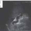
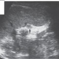
The right adrenal gland lies anterior, medial, and slightly above the upper pole of the right kidney between the renal pole, liver, and vena cava. The left adrenal gland lies anterior and medial to the upper pole of the left kidney between the renal pole and aorta (Figs. 11.1, 11.2).
While the right adrenal gland extends a few centimeters upward behind the vena cava, the left adrenal gland projects only a little above the renal pole and tends to extend downward between the kidney and aorta toward the renal hilum (Figs. 11.1, 11.2).
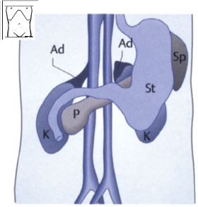
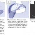
Fig. 11.1 Frontal view of the adrenal glands (Ad). K = kidney, P = pancreas, St = stomach, Sp = spleen.
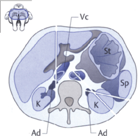
Fig. 11.2 The adrenal glands (Ad) in cross section
Related posts:
Stay updated, free articles. Join our Telegram channel

Full access? Get Clinical Tree


