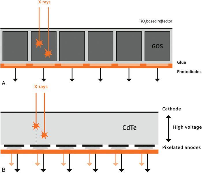
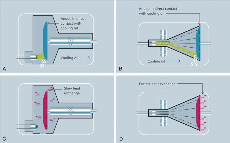
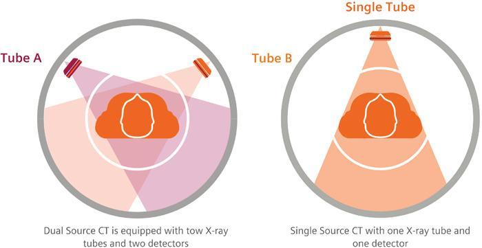
Ashutosh Sharma, Vaibhav Gulati Our future success is directly propotional to our ability To understand, adopt and integrate new technology into our work. Sukant Ratnakar The discovery of X-rays in 1895 by Roentgen revolutionized the field of diagnosis, and the first picture of skeletal system was obtained in 1897; however, the first clinical computed tomography (CT) system was introduced in 1972, a head scanner only after the introduction of computational techniques. Since then, the scientific community and manufacturers have strived to bring about many improvements in the CT technology focussed on getting faster rotation times, more slices, higher spatial resolution, higher temporal resolution, significant X-ray dose reduction, faster image reconstruction times, workflow automation, patient comfort and safety, higher throughput, etc. CT traditionally has been a system for anatomical information; however, introduction of spectral CT has made it possible to get functional status as well. All these technological advancements have facilitated in improving the diagnostic value of the CT and expanding the applications of CT to many clinical fields. Basic components of a CT are X-ray generating system, detectors, collimators and filters and system for reconstruction of images. The technology advancements have been on all fronts and are continuing. Some of the advancements in the various subcomponents of the computed tomography scanner are discussed in the following text. The ceramic material presently used in detectors has made it possible to reduce the detector collimation from 13 mm that was used in the first CT to 0.5 mm now routinely available in the present generation of CT scanners. From single row detectors, we now have multirow detectors in multislice CT. The current generation of solid-state detectors uses a two-step process to transform the incoming X-ray intensity to an electronic signal, with the X-ray first being converted to visible light in the scintillator layer, which is subsequently converted to electric current by a photodiode array, which is digitized in dedicated ASICs (Fig. 1.23.1A). The scintillator layer is made of a ceramic material that is mechanically structured into pixels with septae of finite width separating them to suppress optical crosstalk. Recent research in photon-counting detector technology shows promising advantages, viz.,100% geometric efficiency, no electronic noise, higher spatial resolution and spectral information. At the heart of these detectors are special semiconductor materials that can directly transform X-rays into electric signal pulses. Incoming X-ray quanta generate clouds of free charge carriers in proportion to the energy of the incoming X-ray photon. The charge clouds are transported to anode pixels by a strong electrical field within the detector material, leading to electric currents induced in the pixels. These charge pulses are then converted by highly integrated circuits into voltage pulses measuring a few nanoseconds in duration, which can then be counted digitally (Fig. 1.23.1B). Such detectors will offer remarkable advantages when they become available, allowing not only the counting of each individual photon, but also measuring the energy of each one. They could also allow multienergy CT (real spectral imaging) becoming a possibility without any other technical prerequisites. Possible advantages of photon-counting detectors over conventional detector technology: The X-ray systems in the present generation of the CTs deliver high instantaneous power with high cooling rates of the X-ray tube being still compact enough to be mounted right inside the gantry. The latest generation X-ray tubes have the anode directly in contact with the cooling oil and thus can be efficiently cooled via thermal conduction (Fig. 1.23.2B) unlike the earlier generation of the tubes where the anode cooling is by radiating the heat to the cooling oil (Fig. 1.23.2C). This new generation of tubes deliver very high power up to 120 kW with a very high heat dissipation rate of over 7 MHU/min and above all are compact in size (Fig. 1.23.2D). The conventional tubes have bulky anode to facilitate high heat storage in the anode. This also results in large-sized tube enclosure. New tube technology with compact size and low weight is an enabler for high rotation speeds, long scans ranges and dual-source technology. This type of tube provides improvements for cardiac imaging and fast high-resolution volume scanning where acquisition speed is crucial, such as in whole-body trauma examinations. High temporal resolution is of critical importance specially for cardiac imaging. One of the solutions to achieve high temporal resolution is to have multiple X-ray sources and detectors in the same gantry. The arrangement for having two X-ray sources and two detectors is known as dual-source technology (DSCT). This DSCT makes it possible to achieve extremely high temporal resolution (hardware driven 66 ms) (Fig. 1.23.3).
1.23: Advancements in CT technology
Introduction
CT detector technology
Photon-counting detectors
X-ray system
Dual-source CT scanners – two tubes and two detectors in a gantry
Advantages of dual-source CT
Cardiac
Stay updated, free articles. Join our Telegram channel

Full access? Get Clinical Tree


