11 Anatomy
The discussions of anatomy and embryology in this chapter are limited essentially to structures that either can be visualized with CT or are necessary for understanding a particular disorder. Specialized textbooks may be consulted for details on the ultrastructural morphology and zonal architecture of the spinal cord, which are outside the scope of the present work.
Bone
The spinal column has a segmental architecture consisting of seven cervical vertebrae, 12 thoracic vertebrae, five lumbar vertebrae, five sacral vertebral segments, and three to five coccygeal segments (Fig. 11.1).
While the total number of vertebral bodies is relatively constant, numerous “junctional anomalies” may be encountered, especially in the lumbosacral region. These anomalies may be associated with a variable number of separate lumbar vertebrae, ranging from four to six, due to the sacralization of a lumbar vertebra or the lumbarization of a thoracic or sacral vertebra. Especially in the preoperative diagnosis of disk herniation, these junctional anomalies can make it difficult to define the affected level accurately. To avoid misunderstandings, consultation with the referring physician is recommended whenever possible. In describing the junctional anomaly, the radiologist should use the term “lowest” or “second lowest level,” as this conforms to the intraoperative assessment of vertebral level as determined by fluoroscopy.
It should be noted that the lateral scout view may appear to show six separate lumbar vertebral bodies. This is caused by “short ribs” on the T12 vertebra, which are distinguishable from transverse processes only by the presence of a joint space in the anteroposterior scout view or plain radiograph. This point should be noted in designating the intervertebral spaces (Fig. 11.2).
Except for the axis and atlas (C1 and C2), the basic structural anatomy of the vertebral bodies is consistent throughout the spine and varies only in its adaptation to regional functional demands. All the vertebrae except for C1 possess an anterior (ventral) vertebral body. The pedicles arise from the posterior (dorsal) side of the vertebral body, and combine with the laminae to form the vertebral arch. The anterior surface of the vertebral arch and the posterior surface of the vertebral body form the boundaries of the bony spinal canal. The costotransverse processes arise from the sides of the vertebral arch; they articulate with the ribs in the thoracic spine and serve basically as sites for muscular attachment in the cervical and lumbar spine. Each vertebral body has a superior and inferior articular process, also arising from the vertebral arch, by which it forms facet joints with the adjacent vertebrae.
The segmental differences between the cervical, thoracic, and lumbar regions of the spine include variations in the size and shape of the bony spinal canal. The shape of the canal along the cervical spine ranges from elliptical to triangular, with the apex of the triangle on the dorsal side. The transverse diameter of the cervical spinal canal is greater than its sagittal diameter. It diminishes rapidly from the craniocervical junction to the C3 level and then remains fairly constant through C7 (Figs. 11.3, 11.4).
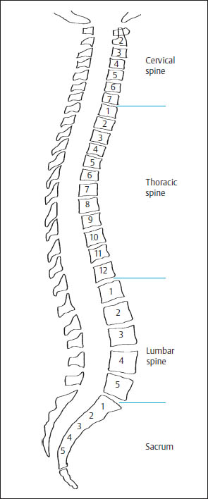
Fig. 11.1 Segmental architecture of the spine.
In the thoracic region, the spinal canal has a rounded cross-section with approximately equal transverse and sagittal diameters. The posterior surface of the thoracic vertebral bodies is concave compared with the cervical and lumbar spine (Figs. 11.3, 11.5).
The lumbar portion of the spinal canal is wider than the thoracic portion and has a somewhat elliptical shape, with a greater transverse than sagittal diameter (Figs. 11.3, 11.6).
The following values are useful guidelines in CT evaluations of spinal canal width (e.g., in patients examined for spinal stenosis):
• The lumbar spinal canal should have an anteroposterior diameter of at least 15 mm.
• The cervical spinal canal below C3 should have an anteroposterior diameter of at least 12 mm.
• The craniocaudal distance between the vertebral arches should be at least 20 mm.
Based on information published in the literature, the following minimum values are useful for excluding spinal stenosis at any level:
• The anteroposterior diameter should measure at least 11.5 mm.
• The interpedicular distance should measure at least 16 mm.
• The width of the lateral recess (the area bounded laterally by the pedicle, posteriorly by the superior articular facet, and anteriorly by the posterolateral surface of the vertebral body) should be no less than 3 mm.
• The area of the spinal canal in axial cross-section should be at least 1.45 cm2.
• The ligamenta flava should not exceed 4–5 mm in thickness.
The thickness of the intervertebral disk spaces (i.e., the disk height) diminishes craniocaudally along the cervical spine. The thoracic disk spaces are somewhat thinner, especially in the upper thoracic area, and become increasingly thick from above downward into the lumbar region. The thickest disk space normally occurs at the L4–5 level, and the L5–S1 disk is slightly thinner.
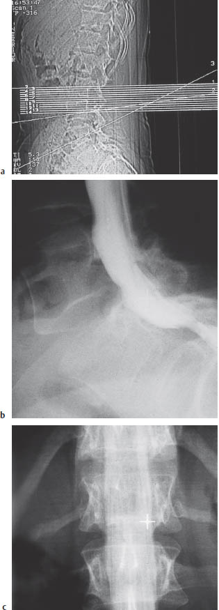
Fig. 11.2a–c Junctional anomaly?
a The lateral scout view appears to show six separate lumbar vertebral bodies, apparently due to the lumbarization of S1.
b, c The lateral myelogram demonstrates an L7-S1 herniated disk. The anteroposterior view shows that the presumed transverse processes of “L1” are actually short ribs. Thus, the normal segmental architecture and the segmental origin of the spinal nerves are preserved, and this must be taken into account when interpreting the images.
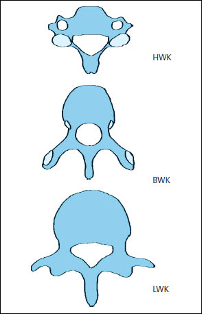
Fig. 11.3 Varying shape of the spinal canal in the cervical, thoracic, and lumbar vertebrae.
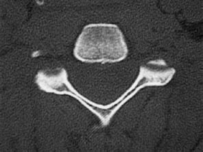
Fig. 11.4 Typical CT appearance of the cervical spinal canal. Triangular cross-section.
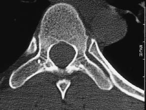
Fig. 11.5 Typical CT appearance of the thoracic spinal canal. Rounded cross-section.
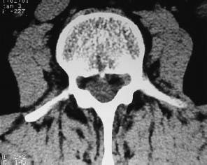
Fig. 11.6 Typical CT appearance of the lumbar spinal canal. Elliptical cross-section.
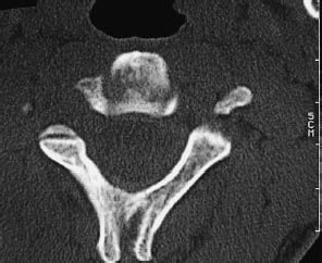
Fig. 11.7 The spinous processes of C2 to C6 have a variable bifid appearance.
Segmental peculiarities of the cervical spine. The vertebral artery and a bundle of small accompanying vertebral veins runs from C6 to C1through the transverse foramina, a series of openings in the transverse processes measuring 5–7 mm in diameter. The foramina may be congenitally open on the posterior, lateral, or anterior side. In another variant, the vertebral artery may pass through a bony “arcuate foramen” over the vertebral artery groove on the arch of the atlas before it enters the foramen magnum.
Usually, the spinous processes of C2 through C6 are bifid in varying degrees (Fig. 11.7).
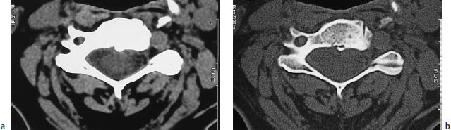
The lateral borders of the C3through C7 vertebral bodies each have a superiorly directed uncinate process that articulates with, and constrains the motion of, the vertebral body above it. In degenerative diseases, this process is often a nidus for appositional new bone growth, which can narrow the neuroforamina and exert pressure on the exiting spinal nerve (Figs. 11.8, 11.9).
The C7 vertebra (nuchal tubercle) is often called the “vertebra prominens” owing to the great length of its spinous process.
Segmental peculiarities of the thoracic spine. Each thoracic vertebra has a small facet on its superior border, just anterior to the pedicle, and on its inferior border for articulation with the heads of the ribs. Another facet on the lateral portion of the transverse processes articulates with the tubercles of the ribs.
Segmental peculiarities of the lumbar spine. In place of costal facets, the lumbar vertebrae have well-developed transverse processes, or costal processes, which represent rudimentary ribs and give attachment to muscles (Figs. 11.6, 11.10).
The atlas (C1) is the only vertebra that lacks an anterior body, consisting basically of a bony ring. This configuration allows for two essential movements at the atlas level. Each lateral mass of the atlas has a superior facet that articulates with the corresponding occipital condyle at the atlanto-occipital joint, which is responsible for nodding movements of the head. Inferiorly, the atlas transmits the load of the head and a portion of the nodding movement through the atlantoaxial joint to the second cervical vertebra (Fig. 11.11).
The ring-like shape of the atlas is particularly favorable for pivoting movements at the atlantoaxial joint, where the dens of the axis articulates with the dental facet on the anterior arch of the atlas (Figs. 11.11, 11.12). The transverse ligament of the atlas retains the dens behind the anterior arch and covers it completely (see Fig. 11.17).
Related posts:
Stay updated, free articles. Join our Telegram channel

Full access? Get Clinical Tree


