Anatomic knowledge is the foundation of clinical phlebology, being crucial for the management of venous disorders in the lower extremity. Our understanding of the morphology and pathophysiology of chronic venous insufficiency and varicose veins has significantly improved in recent years, which made the traditional anatomical nomenclature of the official Terminologia Anatomica 1 somewhat deficient. Accurate terminology of the veins in the lower limb became a challenge for phlebologists given that imprecise identification of veins in the lower limb remained an important source of confusion. The use of inaccurate nomenclature can make the exchange of information in clinical literature difficult and lead to inappropriate management of venous diseases. For instance, the “superficial femoral vein,” which is the main deep vein in the thigh, could be erroneously considered as a vessel of the superficial venous system 2 and abbreviations such as “LSV” could be interpreted as either the “long” or the “lesser” saphenous vein. 3 In addition, the indiscriminate use of eponyms that were not part of the official anatomic terminology could lead to further misconception and confusion. For this reason, in 2001, an International Interdisciplinary Committee was designated to revise the nomenclature of the lower extremity venous system and update the terminology where clinically relevant. 4, 5 This consensus document, stimulated by the need for revision and extension of the Terminologia Anatomica, offered an internationally acceptable venous anatomic terminology for the lower extremity venous system, which is adequate for both anatomists and clinicians.
The terminology and definitions used in this textbook conform to the newest revision of the nomenclature. Therefore, discontinued names such as “superficial femoral vein,” “long or greater saphenous vein,” and “short or lesser saphenous vein” are replaced by their respective accurate terms: “femoral vein,” “great saphenous vein,” and “small saphenous vein,” respectively. Eponyms are discouraged in publications and only a few well-known names that have been retained, such as “Giacomini’s vein,” are mentioned in this chapter. The official Latin terms are written in italics.
1.2 General Considerations
The venules are considered the first part of the venous system, and receive blood from the capillaries ( ▶ Fig. 1.1). These small vascular structures measure 20 µm in diameter, and consist of endothelial cells surrounded by collagen fibers. As the diameter of the venules increases, smooth muscle cells start to appear within the fibrous sheath, and at a diameter of 200 µm, the muscle layer becomes well defined. When the vessel achieves a clinically visible size, the vein is composed of three distinct layers: endothelial lining (tunica intima); smooth muscle cells (tunica media); and collagen fibers (tunica adventitia) ( ▶ Fig. 1.2). The composition of the vein wall is variable, and the muscular content of the media changes depending on the location and on the hydrostatic pressure of the vessel. Microscopic valves in small veins and venules consist of delicate connective tissue lined with endothelial cells; and valves in larger veins are composed of collagen and smooth muscle covered by endothelium. 6, 7, 8, 9, 10, 11
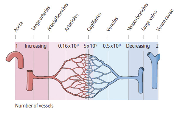
Fig. 1.1 Scheme of the peripheral systemic circulation.
(Reproduced with permission from Schuenke M, Schulte E, Schumacher U. General Anatomy and Musculoskeletal System. Stuttgart: Thieme; 2010. Illustration by Karl Wesker.)
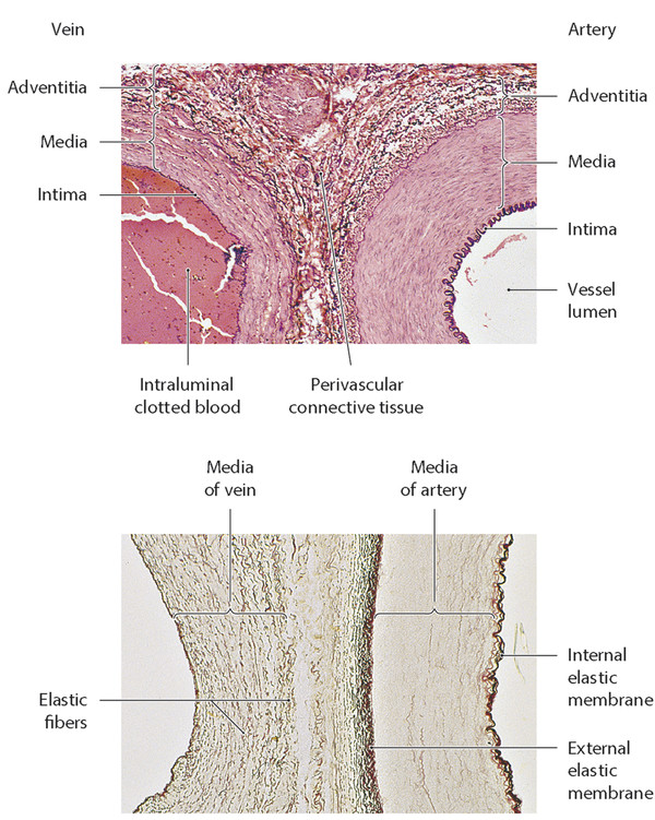
Fig. 1.2 Wall structure of arteries and veins.
(Reproduced with permission from Schuenke M, Schulte E, Schumacher U. General Anatomy and Musculoskeletal System. Stuttgart: Thieme; 2010.)
The anatomy of the lower extremity venous system is extremely variable. As a general rule, the veins of the lower limb are divided into three independent but interconnected systems: the deep venous system, the superficial venous system, and the perforating veins. The deep venous system is located below the muscular fascia, while the superficial venous system lies in the subcutaneous tissue between the muscle fascia and the dermis. The perforating veins are a communication between the superficial and deep venous systems, while communicating veins interconnect with other vessels within the same compartment. 4
1.3 Deep Veins
The deep veins in the lower extremity are located within the deep compartment delineated by the muscle fascia, and accompany the corresponding arteries and their branches. They contain a large number of valves and are regularly doubled as high as the level of the popliteal vein. 12, 13, 14 The structure of the deep venous system of the lower extremity is presented in ▶ Fig. 1.3, with its nomenclature summarized in ▶ Table 1.1.
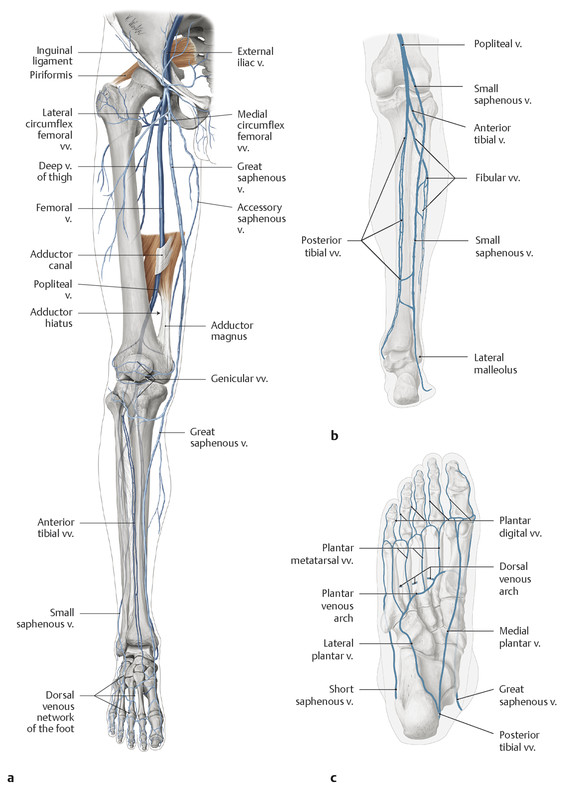
Fig. 1.3 Deep and superficial venous systems of the lower extremity.
(Reproduced with permission from Gilroy AM, MacPherson BR, Ross LM, Schuenke M, Schulte E, Schumacher U. Atlas of Anatomy. 2nd ed. New York, NY: Thieme; 2012. Illustration by Karl Wesker and Marcus Voll.)
Nomenclature | Official Latin term |
Medial plantar veins | Venae plantares mediales |
Lateral plantar veins | Venae plantares laterales |
Deep plantar venous arch | Arcus venosus plantaris profundus |
Deep plantar metatarsal veins | Venae metatarsales plantares profundae |
Deep dorsal metatarsal veins | Venae metatarsales dorsales profundae |
Deep plantar digital veins | Venae digitales plantares profundae |
Deep dorsal digital veins | Venae digitales dorsales profundae |
Pedal veins | Venae dorsales pedis |
Posterior tibial veins | Venae tibiales posteriores |
Anterior tibial veins | Venae tibiales anteriores |
Fibular veins | Venae fibulares |
Genicular venous plexus | Plexus venosus genicularis |
Soleal veins | Venae soleales |
Gastrocnemius veins | Venae gastrocnemii |
Popliteal vein | Vena popliteal |
Common femoral vein | Vena femoralis communis |
Femoral vein | Vena femoralis |
Profunda femoris vein | Vena profunda femoris |
Medial circumflex femoral veins | Venae circumflexae femoris mediales |
Lateral circumflex femoral veins | Venae circumflexae femoris laterales |
Deep femoral communicating veins | Venae comitantes arteriarum perforantium |
Sciatic vein | Vena comitans nervi ischiadici |
1.3.1 Deep Veins of the Foot
The deep veins of the foot are the medial plantar veins (venae plantares mediales), the lateral plantar veins (venae plantares laterales), the deep plantar venous arch (arcus venosus plantaris profundus), the deep plantar metatarsal veins (venae metatarsales plantares profundae), the deep dorsal metatarsal veins (venae metartasales dorsales profundae), the deep plantar digital veins (venae digitales plantares profundae), the deep dorsal digital veins (venae digitales dorsales profundae), and the pedal veins (venae dorsales pedis) ( ▶ Fig. 1.3). The deep plantar digital veins drain the plexuses on the plantar aspect of the toes, communicate with the deep dorsal digital veins, and join to form four deep plantar metatarsal veins running inside the metatarsal space. At the dorsal aspect of the foot, the deep dorsal digital veins and deep dorsal metatarsal veins are arranged in a similar manner. These plantar and dorsal metatarsal veins communicate and join to form the deep plantar venous arch and the pedal veins, respectively. The pedal veins run on both sides of the dorsalis pedis artery, together with the deep fibular nerve. Different from the veins of the leg and the thigh, the valves of veins of the foot are oriented to allow the blood flow from the deep to the superficial veins. 4, 13
1.3.2 Deep Veins of the Leg
The deep veins of the leg are: the posterior tibial veins (venae tibiales posteriores), the anterior tibial veins (venae tibiales anteriores), the fibular veins (venae fibulares), the genicular venous plexus (plexus venosus genicularis), the soleal veins (venae soleales), the gastrocnemius veins (venae gastrocnemii), and the popliteal vein (vena poplitea) ( ▶ Fig. 1.3). The posterior tibial veins accompany the posterior tibial artery and receive tributaries from the posterior group of calf muscles, the venous plexus inside the soleal muscle or soleal veins, the perforator veins, and the fibular vein. These veins contain from 8 to 19 valves. The anterior tibial veins are a proximal continuation of the pedal veins after passing under the inferior extensor retinaculum; they accompany the anterior tibial artery and merge with posterior tibial veins to form the popliteal vein. These veins contain from 8 to 11 valves. The fibular veins travel along the corresponding artery and receive tributaries from the soleal and perforator veins before draining into the posterior tibial vein in the proximal calf. These veins contain from 8 to 11 valves. The gastrocnemius veins are situated within the medial and lateral gastrocnemius muscles and often merge into a common trunk emptying directly into the popliteal vein just distal to the small saphenous vein opening or into the saphenopopliteal junction. The soleal veins are situated in a deeper plane and drain the soleal muscle into either the posterior tibial veins or the fibular veins. The genicular venous plexus, a complex system of interconnected vessels, collects blood from the knee region in the popliteal fossa into the popliteal vein. The popliteal vein ascends through the popliteal fossa receiving several tributaries as described earlier and becomes the femoral vein after passing the adductor hiatus within the insertion of the adductor magnus muscle. The popliteal vein contains from one to four valves and can be duplicated in 5 to 20% of the cases. 4, 13, 14, 15, 16
1.3.3 Deep Veins of the Thigh
The deep veins of the thigh are the common femoral vein (vena femoralis communis), the femoral vein (vena femoralis), the profunda femoris vein (vena profunda femoris), the medial circumflex femoral veins (venae circumflexae femoris mediales), the lateral circumflex femoral veins (venae circumflexae femoris laterales), the deep femoral communicating veins (venae comitantes arteriarum perforantium), and the sciatic vein (vena comitans nervi ischiadici) ( ▶ Fig. 1.3). The femoral vein, previously named with the now unauthorized term “superficial femoral vein,” 2 originates at the superior margin of the popliteal fossa as a direct continuation of the popliteal vein. The vessel courses in the femoral canal and can appear duplicated in up to 46% of cases, isolated or in combination with popliteal vein duplications. Anatomic variations, duplications, and truncular venous malformations are frequently bilateral. The femoral vein usually contains from three to five valves. 13, 14, 17, 18 The profunda femoris vein or deep femoral vein originates as a confluence of smaller muscular veins draining the posterior and lateral thigh and features several valves. The abandoned term “deep vein of the thigh” is nonspecific and misleading and should not be used. 4, 13 The deep femoral communicating veins, formerly named “perforating veins,” accompany the perforating arteries that originate from the deep femoral artery and pierce the adductor muscles to enter the posterior compartment of the thigh. The denomination “perforating,” now reserved for veins that connect to both the superficial and deep venous systems, does not apply to these vessels because they never leave the deep compartment. 4, 13 The medial and lateral circumflex femoral veins drain the thigh muscles and the hip joint frequently into the femoral vein or the common femoral vein, and rarely into the profunda femoris vein. 13 The common femoral vein originates from the confluence of the femoral vein and the profunda femoris vein which is usually situated from 1 to 3 cm distal to the femoral artery bifurcation or 4 to 12 cm distal to the inguinal ligament. The vein is situated within the iliopectineal fossa and becomes the external iliac vein, after passing under the inguinal ligament and entering the pelvis. It only contains one valve, the suprasaphenic valve, which is present in around 80% of cases, located proximally to the saphenous opening, and protects the lower extremity superficial venous system from the elevated intra-abdominal pressures. 4, 13, 14 In 2009, the term sciatic vein (vena ischiadica) was replaced by axial vein of the lower limb (vena axialis membri inferioris) in the Terminologia Embryologica to name the main trunk of the primordial deep venous system. 13 It is located in the posterior portion of the lower extremity following the course of the sciatic nerve, and may persist as an important collateral pathway for the femoral vein. 13, 19 The complete form of the persistent sciatic vein (vena comitans nervi ischiadici) originates from the popliteal vein or tributaries, extends through the dorsal aspect of the thigh and buttock, and terminates into the internal iliac vein. There are two different variations from the complete form: the proximal persistent sciatic vein and the distal persistent sciatic vein. The proximal persistent sciatic vein originates from the posterior superior aspect of the thigh and continues into the pelvis; and the distal persistent sciatic vein originates in posterior inferior aspect of the thigh, terminating into the profunda femoris vein. 13, 19
1.4 Superficial Veins
The superficial veins of the lower extremity are located in the superficial compartment, within the subcutaneous tissue between the dermis and the muscular fascia. The two most important superficial veins are the great saphenous vein (vena saphena magna) and the small saphenous vein (vena saphena parva). In the consensus document of 2002, the terms “great” and “small” were chosen for the nomenclature of the saphenous veins to avoid confusion when abbreviations are used. 4 The saphenous veins are situated in a narrow anatomic space inside the superficial compartment, denominated the saphenous compartment (compartimentum saphenum). The saphenous compartment is bordered superficially by the saphenous fascia and deeply by the muscular fascia, and it contains the saphenous vein accompanied by the saphenous nerve. As a general rule, the accessory saphenous veins (venae saphena accessoriae) are situated outside this compartment, close to the dermis; however, the anterior accessory of the great saphenous vein at the proximal thigh courses deeply in the superficial compartment anteriorly to the great saphenous vein. 4, 5 The structure of the superficial venous system of the lower extremity is presented in ▶ Fig. 1.4 and ▶ Fig. 1.5, with the nomenclature summarized in ▶ Table 1.2.
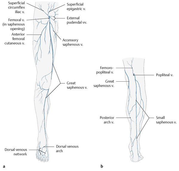
Fig. 1.4 The superficial venous system.
(Reproduced with permission from Gilroy AM, MacPherson BR, Ross LM, Schuenke M, Schulte E, Schumacher U. Atlas of Anatomy. 2nd ed. New York, NY: Thieme; 2012. Illustration by Karl Wesker and Marcus Voll.)
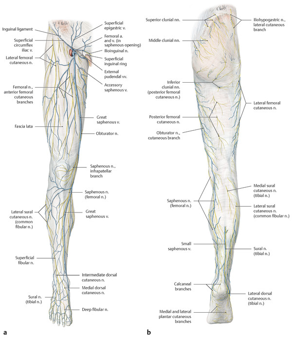
Fig. 1.5 Superficial cutaneous veins and nerves of the right lower limb.
(Reproduced with permission from Gilroy AM, MacPherson BR, Ross LM, Schuenke M, Schulte E, Schumacher U. Atlas of Anatomy. 2nd ed. New York, NY: Thieme; 2012. Illustration by Karl Wesker and Marcus Voll.)
Nomenclature | Official Latin term |
Great saphenous vein | Vena saphena magna |
Superficial epigastric vein | Vena epigastrica superficialis |
Superficial circumflex iliac vein | Vena circumflexa ilium superficialis |
Superficial external pudendal vein | Vena pudenda externa superficialis |
Anterior accessory saphenous vein | Venae saphena magna accessoria anterior |
Posterior accessory saphenous vein | Venae saphena magna accessoria posterior |
Superficial accessory saphenous vein | Vena saphena magna accessoria superficialis |
Anterior thigh circumflex vein | Vena circumflexa femoris anterior |
Posterior thigh circumflex vein | Vena circumflexa femoris posterior |
Small saphenous vein | Vena saphena parva |
Cranial extension of the small saphenous vein | Extensio cranialis vena saphena parva |
Giacomini vein | Vena Giacomini |
Superficial accessory small saphenous vein | Vena saphena parva accessoria superficialis |
Lateral venous system | Systema venosa lateralis membri inferioris |
The veins of the dermis are arranged into two principal horizontal plexuses: the subpapillary venous plexus (plexus venosus subpapillaris) and the reticular venous plexus or deep dermal venous plexus (plexus venosus dermalis profundus). The subcutaneous tissue contains the subcutaneous venous plexus (plexus venosus subcutaneus), which drains into the superficial veins: the saphenous veins and their accessories, collaterals, and tributaries. 20, 21
1.4.1 The Great Saphenous Vein (Vena Saphena Magna)
The great saphenous vein (vena saphena magna), abbreviated as GSV, is the longest vein in the human body, starting at the medial side of the foot and draining into the common femoral vein at inguinal region, through the saphenofemoral junction. It passes anterior to the medial malleolus together with the saphenous nerve and ascends on the medial side of the leg. At the level of the knee, the great saphenous vein courses more posteriorly on the medial aspect of the joint away from the saphenous nerve, which is situated deeper in the thigh. The great saphenous vein then continues to travel alone on the medial side of the thigh, in the superficial compartment, crosses through the saphenous opening (former fossa ovalis), and terminates into the common femoral vein (vena femoralis communis) in the femoral canal ( ▶ Fig. 1.4, ▶ Fig. 1.5). The average diameter of a normal great saphenous vein is 3 to 4 mm, and the vessel features from 10 to 20 valves. 20 The great saphenous vein receives multiple tributaries along its course, and most of these vessels are located superficially outside the saphenous compartment. A variable number of perforator veins connect the GSV to the femoral (vena femoralis), posterior tibial (venae tibiales posteriores), gastrocnemius (venae gastrocnemii), and soleal veins (venae soleales) in the deep venous system.
The term confluence of superficial inguinal veins (confluens venosus subinguinalis) corresponds to the veins of the saphenofemoral junction (junction saphenofemoralis) and is applied to the most proximal segment of the great saphenous vein, close to the common femoral ( ▶ Fig. 1.6). These veins are located between the terminal valve (valvula terminalis), which is the last valve of the GSV, and the preterminal valve (valvula preterminalis), which is situated 3 to 5 cm distally in the superior aspect of the thigh. From the anatomic concept, the saphenofemoral junction corresponds only to the saphenous opening into the femoral vein with the terminal valve. However, in a broader functional concept, the saphenofemoral junction is bounded proximally by the suprasaphenic valve in the common femoral vein and distally by the preterminal valve in the great saphenous vein, and by the infrasaphenic valve in the common femoral vein. This anatomic–functional concept also includes the terminations of the following tributaries that join the saphenofemoral junction: the superficial epigastric vein (vena epigastrica superficialis); the superficial circumflex iliac vein (vena circumflexailium superficialis); the superficial external pudendal vein (vena pudenda externa superficialis); the anterior and posterior accessory saphenous veins (venae saphena magna accessoria anterior et posterior); and the anterior and posterior thigh circumflex veins (vena circumflexa femoris anterior et posterior). The saphenofemoral junction may be correctly abbreviated as SFJ. 4, 5, 20, 22, 23
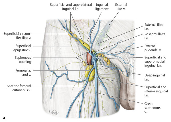
Fig. 1.6 The saphenofemoral junction. (continued)
(a—reproduced with permission from Gilroy AM, MacPherson BR, Ross LM, Schuenke M, Schulte E, Schumacher U. Atlas of Anatomy. 2nd ed. New York, NY: Thieme; 2012. Illustration by Karl Wesker. b—reproduced with permission from Platzer W. Color of Human Anatomy, Volume 1: Locomotor System. Stuttgart: Thieme; 2013. Illustration by Gerhard Spitzer.)
Stay updated, free articles. Join our Telegram channel

Full access? Get Clinical Tree


