Anatomy
1.1 Introduction
The brain is an extremely complex structure with a seemingly random arrangement of ridges (gyri) separated by grooves (sulci). The interior of the brain is composed of structures with a high cellular density (gray matter) in addition to structures with a high fiber density (white matter). When these structures are examined using a sectional imaging modality, they are rarely shown to their full extent. Usually they are visible only in a sectional view, occasionally appearing at multiple sites within the same image. This makes it difficult to appreciate the three-dimensional anatomy of brain structures on sectional images. With magnetic resonance imaging (MRI), this difficulty is compounded by the fact that different sequences produce different tissue contrast, and the selection of the imaging plane is a key factor in determining whether or not an anatomic structure or lesion can be localized.
But that is precisely what is expected of radiologists. Findings that are cloaked in generalities with frequent use of the word “region” will not be taken seriously by the referring neurologist, and rightly so. Radiologists whose oral or written report shows that they are not thoroughly familiar with the region that they are describing and evaluating for pathologic changes do not really justify their existence as a profession. We do not need radiologists to tell us that MRI shows “a mass” or “a hemorrhage” located “in the region of the posterior cranial fossa,” for example. The precise description of a lesion must be accompanied by accurate localization, which often opens the way to making a diagnosis and determining the functional significance of a lesion and the best surgical approach.
Note
The accurate localization of a lesion is the first step on the road to diagnosis.
The outstanding ability of MRI to reveal structural details obliges the radiologist to be at least as good as the technology and to name and classify all brain structures that are visible on the images and can be identified by their location. This chapter is intended to aid in that process. We shall describe landmarks and key structures that facilitate orientation in the superficial and deep regions of the brain. We shall also explore the anatomy of the 12 pairs of cranial nerves and anatomic variants without pathologic significance, relying mainly on standard MRI sequences—mostly T1-weighted (T1w) images—owing to their importance in routine clinical imaging.
1.2 Brain Structures
The brain consists of three main parts:
Cerebrum.
Cerebellum.
Brainstem.
1.2.1 Cerebrum
The cerebrum is subdivided into the following lobes:
Frontal lobe.
Temporal lobe.
Parietal lobe.
Occipital lobe.
Most of the cerebral lobes are delineated by sulci. The central sulcus, for example, divides the frontal lobe from the parietal lobe ( ▶ Fig. 1.1). Thus, the precentral gyrus is part of the frontal lobe while the postcentral gyrus is part of the parietal lobe. The boundary between the occipital lobe and parietal lobe is clearly marked by the parieto-occipital sulcus ( ▶ Fig. 1.2). It is more difficult to locate the boundary between the occipital and temporal lobes on the lateral cerebral surface because they are not separated by a sulcus. An imaginary curved line drawn upward and backward from a small notch at the inferior edge of the inferior temporal gyrus, the temporo-occipital incisure, will roughly define the boundary of the occipital lobe. By contrast, the frontal and temporal lobes are clearly separated from each other by the large sylvian fissure. Not visible on the lateral surface is the insula, which consists of three short and two long gyri, predominantly vertically directed (insular gyri, ▶ Fig. 1.3). The three segments of the frontal, parietal, and temporal lobes that cover the insula make up the operculum (Latin: “little lid”). Accordingly, the three segments are called the frontal operculum, temporal operculum, and frontoparietal operculum. Removing the “little lid” would expose the underlying insular gyri ( ▶ Fig. 1.4).

Fig. 1.1 Structures of the cerebrum.
1 = Middle frontal gyrus
2 = Superior frontal gyrus
3 = Precentral gyrus
4 = “Hand knob”
5 = Central sulcus
6 = Postcentral gyrus
7 = Postcentral sulcus
8 = Superior parietal lobule

Fig. 1.2 Structures of the cerebrum.
1 = Cingulate gyrus
2 = Cingulate sulcus
3 = Central sulcus
4 = Pars marginalis
5 = Precuneus
6 = Parieto-occipital sulcus
7 = Cuneus
8 = Calcarine sulcus
9 = Lingual gyrus
10 = Fornix
11 = Pons
12 = Optic nerve, optic tract
13 = Oculomotor nerve (in section)

Fig. 1.3 Structures of the cerebrum.
1 = Middle frontal gyrus
2 = Transverse temporal gyrus
3 = Insular gyri
4 = Temporal pole
5 = Lateral occipitotemporal gyrus
6 = Cerebellum

Fig. 1.4 Structures of the cerebrum.
1 = Head of caudate nucleus
2 = Putamen
3 = Insular gyri
4 = Column of fornix
5 = Mammillothalamic tract
6 = Third ventricle
7 = Superior colliculus
8 = Anterior commissure
9 = Posterior commissure
1.2.2 Cerebellum
The two hemispheres of the cerebellum are clearly distinguished from the cerebellar vermis by their different surface gyral patterns. The junction of each hemisphere with the vermis is called the pars intermedia.
1.2.3 Brainstem
The brainstem has the following parts, from below upward:
Medulla oblongata ( ▶ Fig. 1.5).
Pons ( ▶ Fig. 1.6, see also ▶ Fig. 1.5).
Midbrain ( ▶ Fig. 1.7):
Two cerebral peduncles (cerebral crura, ▶ Fig. 1.8).
Tegmentum.
Lamina tecti (quadrigeminal plate).
The base of the pons bulges anteriorly toward the clivus and basilar artery. This feature identifies the pons and clearly distinguishes it from the medulla oblongata and midbrain.

Fig. 1.5 Structures of the brainstem.
1 = Sulcus of corpus callosum
2 = Fornix
3 = superior sagittal sinus
4 = Posterior commissure
5 = Pineal gland
6 = Straight sinus
7 = Quadrigeminal plate, lamina tecti
8 = Superior medullary velum
9 = Fastigium
10 = Median aperture (foramen of Magendie)
11 = Genu of corpus callosum
12 = Anterior commissure
13 = Mammillary body
14 = Optic chiasm
15 = Pituitary stalk
16 = Pons
17 = Medulla oblongata

Fig. 1.6 Structures of the brainstem.
1 = Optic nerve
2 = Pituitary stalk
3 = Oculomotor nerve
4 = Inferior temporal gyrus
5 = Pons
6 = Temporal horn
7 = Basilar artery
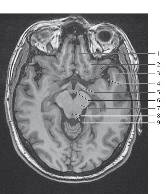
Fig. 1.7 Structures of the brainstem.
1 = Gyrus rectus
2 = Orbital gyri
3 = Temporal pole
4 = Sylvian fissure
5 = Optic tract
6 = Mammillary body
7 = Cerebral crus
8 = Aqueduct
9 = Quadrigeminal plate, lamina tecti, inferior colliculus

Fig. 1.8 Structures of the brainstem.
1 = Anterior orbital gyrus
2 = Olfactory groove
3 = Optic nerve
4 = Optic chiasm
5 = Amygdala
6 = Temporal horn
7 = Cerebral crus
8 = Parahippocampal gyrus
9 = Vermis
10 = Interpeduncular fossa
1.2.4 Magnetic Resonance Imaging of Brain Structures
The basal cerebral arteries and the large cranial venous sinuses can be identified by their flow-related signal loss on MRI. The cranial nerves are usually visualized only in segments that have sufficient caliber and contrast with their surroundings, such as cerebrospinal fluid (CSF) or fat.
All standard imaging planes have advantages and disadvantages. Images in the axial and coronal planes display the symmetry of brain structures and facilitate the detection of any asymmetries. Sagittal images are best for demonstrating the midline structures and the lateral and medial surfaces of the cerebrum and provide excellent orientation.
Note
The best imaging planes for anatomic localization are generally those that display a structure or lesion in its greatest extent. It is also helpful to acquire slices perpendicular to the primary imaging plane. Some cases may require image acquisition in all three planes or even oblique views: for example, to demonstrate the optic nerve in the sagittal plane.
Imaging the curved, infolded surface of the brain in two-dimensional sections poses a unique challenge. Modern workstations have software that can display curved surfaces in a flat plane. But even with these technical capabilities, it should not be forgotten that every sectional imaging study, regardless of plane selection, carries three-dimensional information in all spatial planes. Structures can be tracked along an axis perpendicular to the imaging plane by scrolling through adjacent slices.
1.3 Brain Surface
Tips and Tricks
Lateral sagittal images are best for recognizing and naming the superficial structures of the brain. The midsagittal image, on the other hand, is a longitudinal section through the whole brain that displays the surface of the longitudinal fissure. The sagittal plane is unequaled for the localization and identification of specific gyri. Because gyral anatomy is more constant in the frontal portion of the brain than posteriorly because of the presence of accessory gyri, it is helpful to proceed in an anterior-to-posterior direction when establishing orientation.
Naidich et al (1997) described a very useful, step-by-step method for identifying the structures of the cerebral convexity and their interrelationships. The first step is to identify the sylvian fissure on lateral sagittal slices that display the convexity of the lower frontal lobe and cut the superficial part of the temporal lobe. The widest part of the sylvian fissure is the posterior horizontal ramus, which is continuous anteriorly with the anterior horizontal ramus and anterior ascending ramus. These two rami plus the surrounding inferior frontal gyrus form an M-shaped feature ( ▶ Fig. 1.9) that is easily recognized on images. It consists of three parts: the vertical anterior gyrus is the pars orbitals, the pointed cross-segment is the pars triangularis, and the vertical posterior gyrus is the pars opercularis. The posterior horizontal ramus of the sylvian fissure splits posteriorly into the posterior ascending and descending rami. The anterior and posterior subcentral sulci can be identified as short upward extensions. The posterior ascending ramus is wider in the non-language-dominant hemisphere of the brain.

Fig. 1.9 Surface of the brain. Lateral sagittal image.
1 = Pars triangularis
2 = Pars opercularis
3 = Posterior ascending ramus
4 = Posterior descending ramus
5 = Posterior subcentral sulcus
6 = Lateral sulcus, posterior horizontal ramus
7 = Anterior subcentral sulcus
8 = Anterior ascending ramus
9 = Anterior horizontal ramus
10 = Pars orbitalis
Above the inferior frontal gyrus (the M shape just described) is the inferior frontal sulcus, which separates the inferior frontal gyrus from the middle frontal gyrus. The middle frontal gyrus has a typical zigzag shape. The inferior frontal sulcus turns downward and forward to form the lower portion of the precentral sulcus, which is the sulcus that posteriorly bounds the pars opercularis of the M-shaped inferior frontal gyrus. The precentral sulcus, which runs upward and backward, is interrupted superiorly at the site where the middle frontal gyrus fuses with the precentral gyrus. The level of the fusion site is variable.
Posterior to the precentral sulcus is the precentral gyrus, which is connected to the postcentral gyrus by a bridge, the subcentral gyrus, near the sylvian fissure. The subcentral gyrus is located between the short anterior and posterior subcentral sulci. The pre- and postcentral gyri are separated by the central sulcus. The postcentral sulcus may be continuous with the intraparietal sulcus. This area may have a variable appearance due to the presence of accessory gyri such as the presupramarginal gyrus or preangular gyrus. The posterior end of the sylvian fissure, the posterior ascending ramus, is bounded superiorly by the horseshoe-shaped supramarginal gyrus ( ▶ Fig. 1.10). Its posterior limb fuses with the superior temporal gyrus. Just posterior to the supramarginal gyrus is a second horseshoe-shaped gyrus, which winds around the superior temporal sulcus and connects the posterior portion of the superior temporal gyrus to the middle temporal gyrus. That structure is the angular gyrus. The anatomy of this area is less constant than in the frontal lobe, however, due to the possible presence of accessory gyri. Above the supramarginal gyrus and angular gyrus is the intraparietal sulcus. It separates the supramarginal and angular gyri, as well as accessory gyri that make up the inferior parietal lobule, from the superior parietal lobule, a group of gyri that are bounded anteriorly and superiorly by the postcentral sulcus and posteriorly and inferiorly by the intraparietal sulcus.
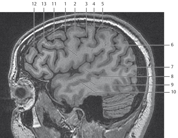
Fig. 1.10 Surface of the brain. Lateral sagittal image.
1 = Precentral sulcus
2 = Precentral gyrus
3 = Central sulcus
4 = Postcentral gyrus
5 = Postcentral sulcus
6 = Supramarginal gyrus
7 = Angular gyrus
8 = Transverse temporal gyrus (Heschl gyrus)
9 = Superior temporal sulcus
10 = Superior temporal gyrus
11 = Subcentral gyrus
12 = Inferior frontal gyrus
13 = Middle frontal gyrus
The transverse temporal gyrus (of Heschl) is clearly visible in lateral sagittal images (see ▶ Fig. 1.10). It is the gyrus that is perched on top of the superior temporal gyrus and protrudes into the sylvian fissure. Behind it, separated by the Heschl sulcus, is the planum temporale, which is larger in the language-dominant hemisphere. The Heschl gyrus may be indented by an intermediate sulcus, changing it from an omega (ω) shape to a heart shape. Reportedly, when this gyrus is viewed in axial section it is usually found in the plane of the interthalamic adhesion but may also be found running obliquely forward in the next higher or lower section. As a landmark for locating the Heschl gyrus in the coronal plane, Yousry et al (1997) describe the section in which the crura of the fornix unite at an acute angle or the section in which the internal acoustic meatus can be seen ( ▶ Fig. 1.11). In most cases the Heschl gyrus also exhibits an omega shape when viewed in the coronal plane.

Fig. 1.11 Surface of the brain. Coronal image.
1 = Trunk of corpus callosum
2 = Fornix
3 = Sylvian fissure
4 = Pons
5 = Trigeminal nerve
6 = Internal acoustic meatus
7 = Transverse temporal gyrus (Heschl gyrus)
The central sulcus is an important landmark because it marks the location of the functionally important pre- and postcentral gyri. Many authors have given special attention to identifying this sulcus. It is most easily identified in high axial slices. An easy way to locate it is to follow the superior frontal gyrus posteriorly until it meets a gyrus that runs forward and downward—the precentral gyrus ( ▶ Fig. 1.12a). At that location, both gyri form an L shape on the left cerebral hemisphere. Often the corresponding sulci, the superior frontal and precentral sulci, also form an L shape ( ▶ Fig. 1.12b). Another way to identify the precentral gyrus is by noting its shape: the precentral gyrus thickens near the midline to form a nodule (see ▶ Fig. 1.1) or double nodule in the hand motor area. This nodule, called the “hand knob,” bulges posteriorly into the central sulcus. The hand motor area appears in sagittal images ( ▶ Fig. 1.13) as a typical hook that points posteriorly. The “hook” appears in the sagittal section that also cuts the insular gyri. Naidich and Brightbill described a more detailed method of locating these central structures based on the midsagittal image and high axial slices. An important landmark is the pars marginalis ( ▶ Fig. 1.14), which is the terminal part of the cingulate sulcus. Viewed in the midsagittal plane (see ▶ Fig. 1.2), the cingulate sulcus accompanies the cingulate gyrus as it rounds the corpus callosum. Just before reaching the splenium it curves away from that path and runs apically, terminating on the brain surface as the pars marginalis. Because the central sulcus does not open into the midline but ends medially at the paracentral lobule (see ▶ Fig. 1.12a), it appears only as a small indentation (see ▶ Fig. 1.2) anterior to the pars marginalis. Posterior to the vertical portion of the cingulate sulcus and pars marginalis is the precuneus, which is bordered on its posterior aspect by the parieto-occipital sulcus. Anterior to the vertical portion of the cingulate sulcus is the paracentral lobule. The relatively small occipital lobe is subdivided by the calcarine sulcus, forming an upper portion called the cuneus.

Fig. 1.12 Surface of the brain. High axial image. (a) Superficial structures. (b) The superior frontal gyrus and precentral gyrus form an L shape (shown in white), as do the superior frontal sulcus and precentral sulcus (shown in blue).
1 = Superior frontal gyrus
2 = Superior frontal sulcus
3 = Middle frontal gyrus
4 = Precentral sulcus
5 = Precentral gyrus
6 = Central sulcus
7 = Postcentral gyrus
8 = Pars marginalis
9 = Paracentral lobule

Fig. 1.13 Surface of the brain. High axial image. The insula marks the location of the hand knob.
1 = Precentral gyrus
2 = Central sulcus with hand knob
3 = Postcentral gyrus

Fig. 1.14 Surface of the brain. High axial image. The pars marginalis is shown in blue.
High axial images (see ▶ Fig. 1.12 and ▶ Fig. 1.14) depict the pars marginalis as a sulcus on both sides of the midline. The sulcus curves forward, creating the appearance of a shallow bowl, with the apical end of the central sulcus pointing into the bowl. Anterior to the pars marginalis, the upper portions of the pre- and postcentral gyri fuse to form the paracentral lobule. A bridge connects the postcentral gyrus to the precuneus and marks the endpoint of the postcentral sulcus in its upper medial portion. In most cases—though not in the images shown here—the postcentral sulcus has a bifid termination with two short arms that bracket the pars marginalis on the anterior and posterior sides.
The occipital lobe is most easily identified on the medial surface of the cerebral hemisphere (see ▶ Fig. 1.2). The conspicuous parieto-occipital sulcus ( ▶ Fig. 1.15) runs almost perpendicular to the tentorium and straight sinus, with all three structures defining the anteroinferior boundary of the occipital lobe. The calcerine sulcus, important for central vision, is most clearly displayed on parasagittal images (see ▶ Fig. 1.2). Its anterior portion runs parallel to the tentorium in this plane, subdividing the occipital lobe into the cuneus above and the medial occipitotemporal gyrus or lingual gyrus below. The occipital lobe should be distinguishable from the posterior temporal lobe on lateral sagittal images by the preoccipital notch. Viewed from below (with the cerebellum removed), there is no visible boundary between the temporal and occipital lobes: the anterior-to-posterior course of the medial and lateral occipitotemporal gyri is uninterrupted ( ▶ Fig. 1.16).

Fig. 1.15 Surface of the brain.
1 = Corpus callosum
2 = Parieto-occipital sulcus
3 = Anterior commissure
4 = Preterminal gyrus
5 = Gyrus rectus
6 = Cingulate gyrus

Fig. 1.16 Surface of the brain.
1 = Parahippocampal gyrus
2 = Collateral sulcus
3 = Lateral occipitotemporal gyrus
4 = Superior temporal gyrus
5 = Middle temporal gyrus
6 = Inferior temporal gyrus
The inferior temporal gyrus can be identified lateral to the occipitotemporal gyrus on both coronal and parasagittal images (see ▶ Fig. 1.16). Just above and lateral to the inferior temporal gyrus are the middle and superior temporal gyri, separated by the superior and inferior temporal sulci. While the lateral occipitotemporal gyrus can be traced almost to the temporal pole on the inferior surface of the temporal lobe, the medial occipitotemporal gyrus—bounded by the collateral sulcus—gives way anteriorly to the parahippocampal gyrus ( ▶ Fig. 1.17, see also ▶ Fig. 1.18), which surrounds the upper brainstem and ambient cistern on both sides. Thus the parahippocampal gyrus forms the most medial surface of the temporal lobe and, together with the cingulate gyrus, forms the peripheral boundary of the limbic system. The dentate gyrus, so called because of its regular indentations ( ▶ Fig. 1.18), borders on the parahippocampal sulcus and collosal sulcus at a deeper concentric level. The dentate gyrus is continuous with the indusium griseum at the splenium of the corpus callosum. The indusium griseum is a thin layer of gray matter on the dorsal surface of the corpus callosum. A thin layer of white matter, the fimbriae, is located above the dentate gyrus at a deeper level in the concentric limbic system. The fimbriae overlie the hippocampus and parahippocampal gyrus and run upward and backward with it, becoming continuous with the crura of the fornix. The crura move closer together and are interconnected by fine, stringlike structures before joining at the midline to form the body of the fornix. The commissure of the fornix has been termed the “psalterium” owing to its harplike appearance. The body of the fornix forms the lower boundary of the septum pellucidum and the roof of the cisterna veli interpositi, located between the fornix and the roof of the third ventricle. Farther anteriorly and inferiorly, the columns of the fornix diverge toward both sides. Some of the fornix fibers then run in front of the anterior commissure while most run behind it and soon reach the mammillary bodies ( ▶ Fig. 1.19).

Fig. 1.17 Surface of the brain. Parahippocampal gyrus. (a) The “crosshairs” mark the location of the parahippocampal gyrus. (b) Parahippocampal gyrus in an axial slice angled parallel to the temporal lobe. (c) Parahippocampal gyrus in the coronal plane.

Fig. 1.18 Surface of the brain.
1 = Dentate gyrus

Fig. 1.19 Surface of the brain.
1 = Fornix (column)
2 = Mammillary body
The hippocampal formation consists of a head, body, and tail:
Head: The head is the most anterior part of the hippocampus. It is aligned transversely and is separated from the more anterior amygdala by the uncinate recess of the temporal horn.
Body: Located just behind and above the head, the body of the hippocampus has a predominantly parasagittal alignment and is bounded laterally and superiorly by the temporal horn of the lateral ventricle.
Tail: The tail of the hippocampus has an almost transverse orientation. It extends around the splenium and is continuous with the indusium griseum.
The internal structure of the hippocampus is best appreciated in sagittal or coronal images. Viewed in parasagittal images, the hippocampal formation appears as a collection of gray matter that is parallel to the temporal horn with a posterosuperior-to-anteroinferior alignment. The fimbriae separate the gray matter from the ependyma of the ventricle. With good optical resolution, the gray matter of the Ammon’s horn can be distinguished from that of the dentate gyrus and subiculum ( ▶ Fig. 1.20). Coronal slices for optimum evaluation of the hippocampus should be angled in a counterclockwise fashion, perpendicular to the surface of the temporal lobe (see ▶ Fig. 1.17c and ▶ Fig. 1.20). The parahippocampal gyrus, bounded laterally by the collateral sulcus, is located below the hippocampal formation and forms the medial boundary of the undersurface of the temporal lobe (see ▶ Fig. 1.17 and ▶ Fig. 1.20). When this gyrus turns back medially it is called the “subiculum,” which forms a connection between the parahippocampal gyrus and Ammon’s horn. The Ammon’s horn suggests premature petrification because, during its development, it curves upward and finally assumes the shape of a coiled snail when viewed from the front. The end of this convolution dips into another U-shaped gyrus, the dentate gyrus. This formation is bounded laterally and superiorly by the temporal horn ependyma of the lateral ventricle (see ▶ Fig. 1.20). The fimbriae appear as a thickened area on the medial surface of the hippocampal formation above the Ammon’s horn. Viewed in the coronal plane, the fimbriae run laterally upward to a tapered point (taenia fimbriae) that marks the boundary between the temporal horn and the transverse fissure of the subarachnoid space, which is infolded from the medial side at this location. With good spatial resolution, the stria terminalis and tail of the caudate nucleus can be identified directly above the temporal horn, which is usually narrow, and over the Ammon’s horn.

Fig. 1.20 Surface of the brain.
1 = Fimbria of hippocampus
2 = Temporal horn
3 = Ammon’s horn
4 = Dentate gyrus
5 = Subiculum
6 = Parahippocampal gyrus
Note
An accurate determination of the size and signal intensity of the hippocampal formation is the basis for diagnosing hippocampal sclerosis.
1.3.1 Illustrative Cases
The following four cases illustrate the principle that MR images must be acquired in at least two planes to establish the true location of a structure or lesion in the brain.
1.3.1.1 Case 1
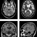
The right-sided tumor in this case appears to be located in the parietal lobe relative to the opposite side ( ▶ Fig. 1.21a). But a sagittal image ( ▶ Fig. 1.21b) reveals the error: the tumor is in the temporal lobe. The large mass has caused marked swelling and enlargement of the temporal lobe with associated compression of the parietal lobe.

Fig. 1.21 Case 1. (a) Axial plane. (b) Sagittal plane.
1.3.1.2 Case 2
This case also illustrates a tumor. On axial MRI ( ▶ Fig. 1.22a), it is unclear whether the tumor is in the frontal or parietal lobe. The coronal image does not aid localization ( ▶ Fig. 1.22b). When a sagittal view is added ( ▶ Fig. 1.22c), it localizes the tumor to the precentral gyrus.

Fig. 1.22 Case 2. (a) Axial plane. (b) Coronal plane. (c) Sagittal plane.
Related posts:

Stay updated, free articles. Join our Telegram channel

Full access? Get Clinical Tree


