Jean Noel Buy1 and Michel Ghossain2
(1)
Service Radiologie, Hopital Hotel-Dieu, Paris, France
(2)
Department of Radiology, Hotel Dieu de France, Beirut, Lebanon
23.1 Anatomy []
23.2.1 The Outer Layer
23.2.2 The Middle Layer
23.2.3 The Inner Layer
23.3.1 Arteries
23.3.2 Veins
23.3.3 Lymphatic Drainage
23.4 Histology []
23.5.1 Ultrasound
23.5.2 MR
23.5.3 Contractions
Abstract
The body of the uterus forms the upper two-thirds of the uterus. It is pear shaped, while the cervix is narrower and is cylindrical in shape. The adult nonpregnant uterus is about 7 cm length, 5 cm in breath, and 3 cm thick.
23.1 Anatomy [1]
The body of the uterus forms the upper two-thirds of the uterus. It is pear shaped, while the cervix is narrower and is cylindrical in shape. The adult nonpregnant uterus is about 7 cm length, 5 cm in breath, and 3 cm thick.
The body of the uterus extends from the fundus at its uppermost part to the cervix inferiorly. Near its upper end, it receives on both sides the uterine tubes. The point of fusion between the uterine tube and body is called the uterine cornu.
Infero-posterior to the cornu is the ovarian ligament. Infero-anterior to the cornu is the round ligament (see Chap. 2). In fact these two structures are in continuity in the embryo and are both derivates from the gubernaculum.
The fundus is covered by peritoneum, which is continuous with that of anterior and posterior surfaces. The lateral margins of the body are convex, and on each side their peritoneum is reflected laterally to form the broad ligament (See Chap. 2).
The peritoneum of the anterior surface is reflected onto the bladder at the uterovesical fold.
The peritoneum of the posterior surface continues down to cover the cervix, and the upper vagina then is reflected back to cover the rectum along the surface of the Douglas.
23.2 Structure of the Myometrium [2]
The body of the uterus from outside to inside has three layers (Fig. 23.1):


Fig. 23.1
Vertical section of the uterine wall close to the fundus. 1 endometrium: 1′′ surface epithelium (small oblique arrow), 1″ glands and stroma, 2 muscularis: 2′ inner layer, 2″ middle layer, 2″ outer layer, 3 serosa: mesothelium and its conjunctive layer (From Testut [2])
1.
Serosa: it comprises mesothelium and its conjunctive-elastic tissue
2.
Muscularis: the myometrium
The muscularis of the body comprises from outside to inside three different layers (Figs. 23.1 and 23.2).
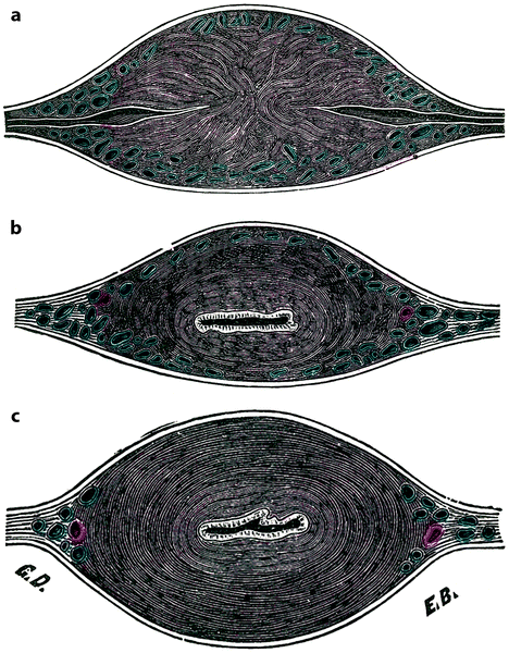

Fig. 23.2
Anatomic constitution of the uterine wall on transverse section of the uterus (the anterior face is at the inferior part of the figure). (a) Through the superior portion of the body, at the level of the tubal orifices. (b) Through the middle part of the body. (c) At the middle part of the cervix (From Testut [2])
23.2.1 The Outer Layer
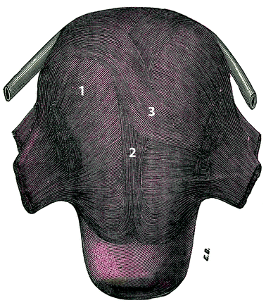
The outer layer (Fig. 23.3) comprises outer longitudinal fibers and immediately beneath them transverse fibers.
Get Clinical Tree app for offline access


Fig. 23.3
Anterior view of the uterus: muscularis outer layer. 1 transverse fibers, 2 longitudinal fibers, 3 fascicle with a Z shape (From Testut [2])
(a)
Longitudinal Fibers. They form a flattened fascicle of 10–25 mm width, which occupies the midline of the uterus on its anterior face, on the fundus, and on the posterior face, with a horseshoe shape. This fascicle goes down a little lower on the posterior surface of the uterus until its middle third of the cervix, while on its anterior surface, it stops at the junction of the body and the cervix.
This fascicle is constituted initially on the anterior face as on the posterior face by fibers initially transverse that arise from the lateral parts of the uterus and then straighten up to become vertical.
When they arrive on the fundus of the uterus, the fibers follow a double direction. Some pass directly from the anterior face onto the posterior face and vice versa. The other ones bend outside to become transverse to make one’s way to the orifices of the tubes. Among these ones, some cross the midline and draw an elongated Z.
(b)
Transverse fibers immediately beneath the previous ones form a regular and continuous plane all over the length of the uterus. On the lateral borders of the uterus:
Some curve to pass from the anterior face to the posterior face of the uterus; they are crossed by numerous arteries and veins forming around them round or elliptical rings.
Stay updated, free articles. Join our Telegram channel

Full access? Get Clinical Tree


