Anatomy in Sagittal MPRs
The images on the next seven pages illustrate the sectional anatomy of the chest and abdomen as it appears in a right-to-left series of sagittal reconstructions.
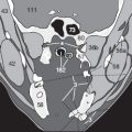
The first sagittal plane ( Fig. 214.1 ) through the far right side of the body shows the oblique muscles of the abdominal wall (28), sections of the ribs (51) with the intercostal muscles (25), and a section of the scapula (53) with the subcostal muscle (18) and infraspinatus muscle (20). The next image in a slightly more medial plane ( Fig. 214.2 ) displays the right lobe of the liver (122) and a section of the ascending colon (142). The numerical labels are keyed to the “Thoracic and Pelvic Diagrams” fold-out key on the back cover flap of this book.


These images ( Figs. 215.1–3 ) display the gallbladder (126), the parenchyma of the right kidney (135) with the renal pelvis (136), and the transverse colon (143). Please note the appearance of thin colon walls, sharply outlined towards surrounding mesenteric fat (2). A blurred appearance of colon walls would suggest either an inflammatory process or an infiltration by a malignant tumor.


Like in transverse sections, you might also come across a subtle partial volume effect of the renal pelvis (136), filled with branches of renal vessels (110, 111) and dark fat (2), which should not be confounded with renal tumors. The more we move from right to left, the smaller the size of subdiaphragmatic hepatic segments (122) will appear ( Figs. 216.1 and 2 ).


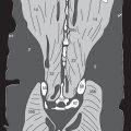
On this page, we approach the paramedian sagittal image level with the inferior vena cava (80) and the abdominal aorta (89). Again, you can see here moderate to severe arteriosclerotic plaques (174) attached to the aortic wall ( Fig. 217.2 ), which narrow or might even occlude the vascular lumen (see Fig. 211.2 , same patient).
Above the diaphragm (30), you can identify the ascending aorta (89a), pulmonary blood vessels (96) and heart chambers (74a-c).


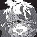
While we move further towards the left paramedian side, the ascending aorta (89a), aortic arch (89b) and descending aorta (89c) come into our field of view, as well as the left ventricle (74d). In Fig. 218.1 , we can identify the celiac trunk (97) with left gastric artery (109) and superior mesenteric artery (106), originating from the suprarenal part of the abdominal aorta (89). In the center of the mesenteric root, we see branches of small mesenteric blood vessels (108) running towards small bowel loops (140) and colon (143), which is partially filled with gas (4 in Fig. 218.2 ).


Further to the left, the stomach (129) with usually thin gastric walls and the left renal pelvis (136) are visualized. Please note that the myocardial wall of the left ventricle (74d in Figs. 219.1–2 ) is significantly thicker compared to right ventricular walls (74b in Fig. 218.2 ). In normal findings, both bowel walls (140, 143) and renal borderlines are sharply defined by adjacent mesenteric fatty tissue (2).


In elder patients, not only obesity, but also benign renal cysts (169) can often be observed ( Fig. 220.1 ) and fat and postinflammatory scar tissue (186) in the lower parts of the pleural cavity, close to the diaphragm (30). Posteriorly, just under the diaphragm, we find the spleen (133), which might appear inhomogeneous in early arterial phases of CM-enhanced images ( Figs. 220.1–3 ), but should be homogenous in late venous perfusion phases (see p. 127).
Related posts:


Stay updated, free articles. Join our Telegram channel

Full access? Get Clinical Tree


