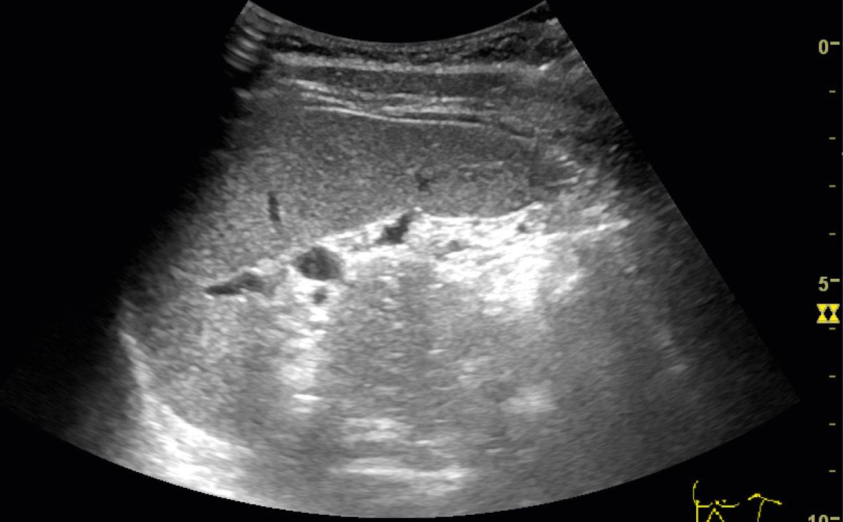
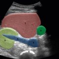
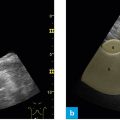
Fig. 1: Position of the probe, intercostal, left flank – spleen and left kidney.
The probe is placed along the left flank, in the intercostal space, at the level of the mid-axillary line. One thus obtains an oblique section in which the probe marker – in this exceptional instance – points away from the investigator. This position is usually in a more dorsal and cranial aspect than one would expect of the spleen.
The following steps are crucial to obtain an image of good quality:
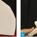
- To obtain an image devoid of artifacts despite positioning the probe in the intercostal space, the probe is rotated until the acoustic shadows of the ribs move to the margin of the image.
- First one observes and measures the spleen alone.
- One intercostal space further in the caudal aspect, one finds a sectional plane for the spleen and the left kidney, or only the left kidney.
Optimal viewing
The following structures should be viewed:
- Spleen
- Hilum of the spleen
- Left kidney
- Possibly the tail of the pancreas