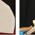
Fig. 1: Direction of probe motion to locate the aortic bifurcation.
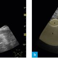
From its position for the pancreas the probe is moved in parallel fashion in caudal direction.
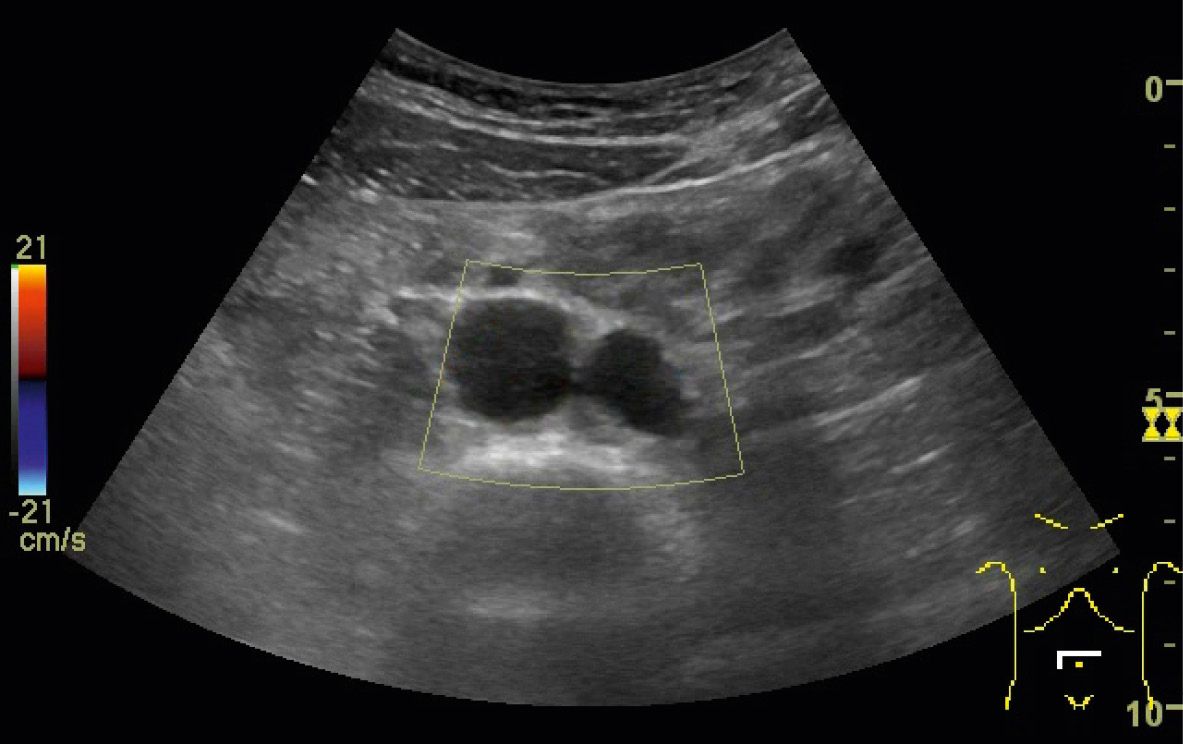
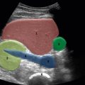
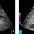
At the umbilical level the aorta becomes more prominent and eventually divides into two large vessels. The aortic bifurcation is seen visible. One may even identify the points of exit as well.
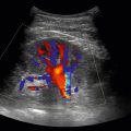
The aortic bifurcation is clearly seen in the majority of patients, and clearly shows the close proximity of important structures at the umbilical level. When the bowel and its acoustic shadow superimpose the bifurcation or the aorta bifurcates at the umbilical level, one must exert sufficient pressure on the probe and achieve appropriate angulation. Color Doppler may help to visualize the bifurcation more clearly.
The following structures should be viewed:
- Aorta
- Aortic bifurcation
- Optional: inferior mesenteric artery