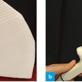
Fig. 1: Position of the probe, subxiphoid, lengthwise – great vessels
The probe is placed lengthwise directly below the xiphoid. One thus obtains a longitudinal view of the great vessels. By performing slight swiveling motions in lateral direction one can also distinguish the aorta and the vena cava. The probe marker points cranially.
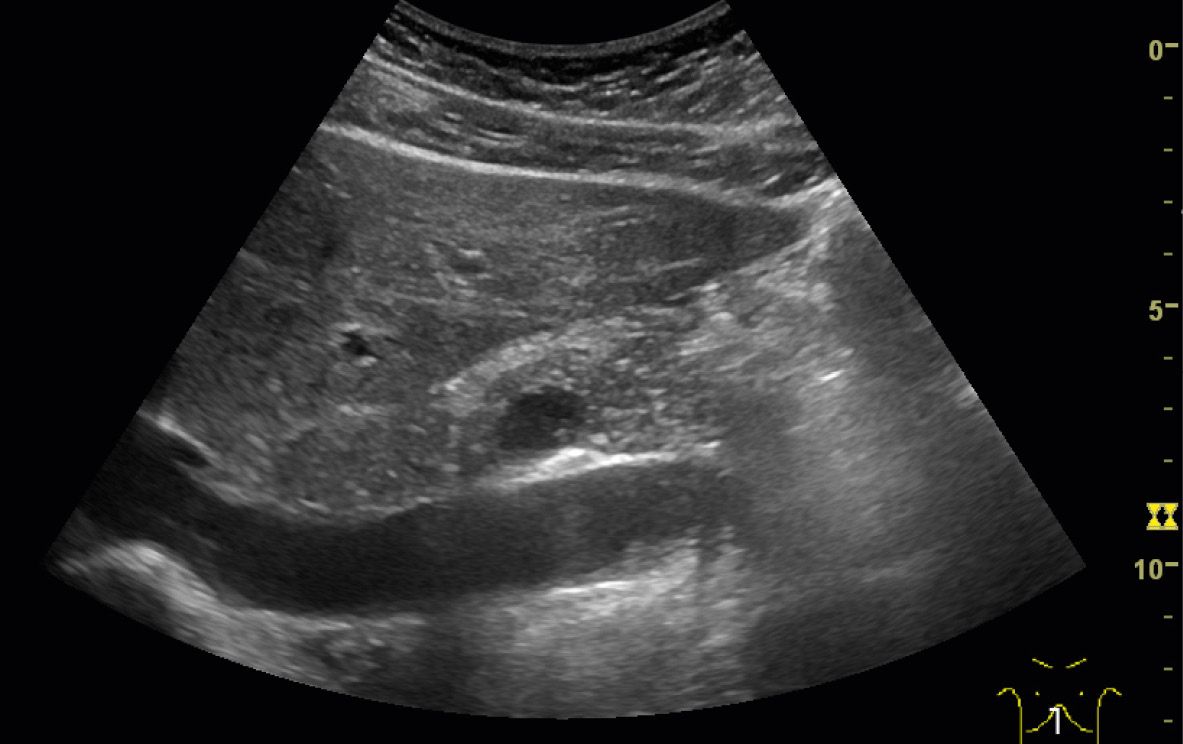
One obtains best results by placing the probe just below the xiphoid. The liver serves as a reliable acoustic window in non-fasting or obese patients. To distinguish the aorta and the vena cava, one may utilize the anatomical position of each or look for the vessels points of exit.
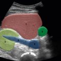
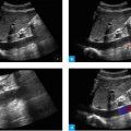
The vena cava is usually seen better – it may be hypoechoic or anechoic and has no inflowing vessels in this region. The aorta is also hypoechoic, but has a washed-out appearance. Two large points of exit can be clearly identified in the distal aspect of the diaphragm: the celiac trunk and the superior mesenteric artery.
Pulsations are no reliable distinguishing feature. The vena cava may mimic pulsations because of venous undulations or wave-like motions.
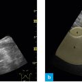
Differentiation of the aorta and the vena cava by their anatomical position and points of exit
The following structures should viewed:
- Vena Cava
- Aorta
- Exit points of arteries:
- Celiac trunk (TC),
- Superior mesenteric artery (SMA)
- Celiac trunk (TC),