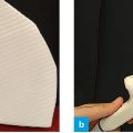
Fig. 1: Position of the probe – oblique subxiphoid – direction of shift for the renal arteries.
To view the pancreas and the superior mesenteric artery the probe is slided caudally parallel to the skin from the transverse view. The marker points to the investigator in the lateral aspect. As bowel gases may superimpose the renal arteries, it may be necessary to make the patient lie on
his/her side.
Visualizing the renal arteries from the anterior position is a challenge during ultrasound investigation of the abdomen. Depending on superimposition due to gases, it may be very difficult to inspect the renal arteries or measure these efficiently. To obtain a clear view one should press away the bowel slightly by exerting sufficient pressure, and the patient should be lying on his/her side so that the bowel shifts on its own to the lateral aspect.
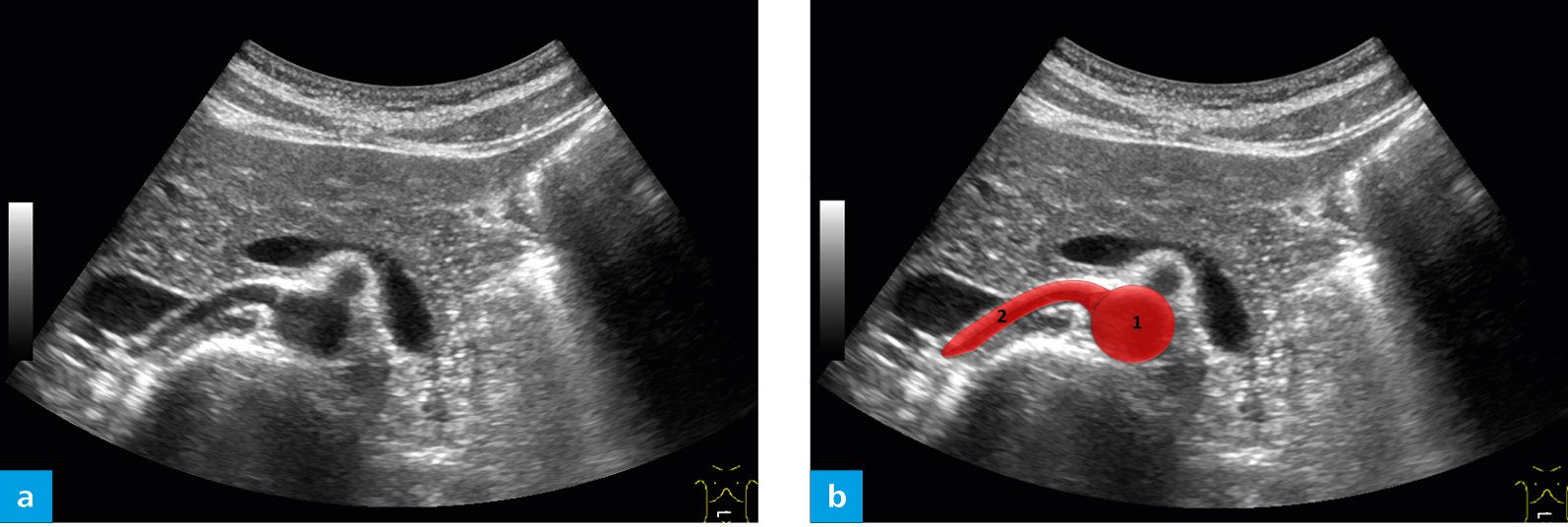
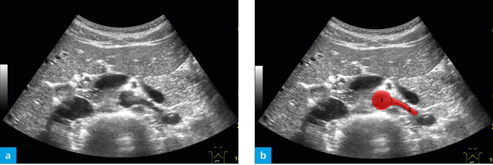
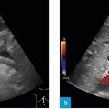
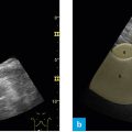
Fig. 2: Ultrasound investigation of the right renal artery on the B-mode image (a); with marking (b) of the aorta (1) and the right renal artery (2).
The right renal artery courses in the dorsal aspect of the vena cava.
Related posts:


Stay updated, free articles. Join our Telegram channel

Full access? Get Clinical Tree


