Fig. 1: Position of the probe, suprapubic, transverse – bladder.
The probe is placed transversely on a line between the two points of the anterior superior iliac spine, in the suprapubic aspect. The probe marker points laterally to the investigator. The advantage of the suprapubic cross-section is that it continues the section used for the aortic bifurcation, and permits better delineation of structures than on the longitudinal section.
The bladder may vary in size, depending on its level of filling. Due to its fluid content the bladder is clearly hypoechoic or anechoic. In addition to the usual anatomical structures one should always view the pouch of Douglas, which is the deepest site in the abdominal cavity. In men this cavity lies between the bladder and the rectum; in women it is between the bladder and the uterus. The following ultrasound images show the male pelvis:
The following structures should be viewed:
- Bladder
- Prostate (man), uterus and ovaries (woman)
- Pouch of Douglas
- Rectum
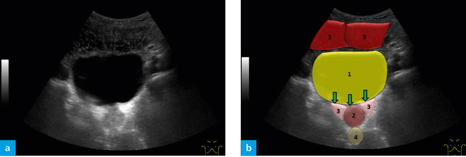
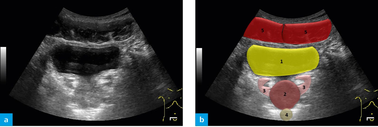
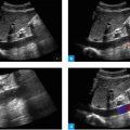
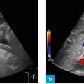
Fig. 2: Ultrasound investigation of a massively filled bladder, B-mode image (a). Bladder (1), prostate (2), the region of the seminal gland (3), rectum (4), muscle bellies of the rectus abdominis muscle (5) (b).
In a massively filled bladder as in Figure 2, the dorsal acoustic shadow is very prominent and may even impair one’s view of the prostate because of excessive echogenicity. In the presence of a massively filled bladder, the prostate and the rectum can be seen only from an image depth of about 10 cm; this additionally impairs resolution on the image.
Related posts:


Stay updated, free articles. Join our Telegram channel

Full access? Get Clinical Tree


