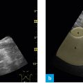
Fig. 1: Position of the probe – right flank.
The probe is placed lengthwise in the right flank, between the anterior and posterior axillary line, directly below the costal arch. The marker points in cranial direction
When the picture is superimposed by the bowel, one moves the probe in parallel fashion further into the dorsal aspect. When ribs cause an unfavorable acoustic shadow, one positions the probe in the intercostal space.
The following structures should be viewed:
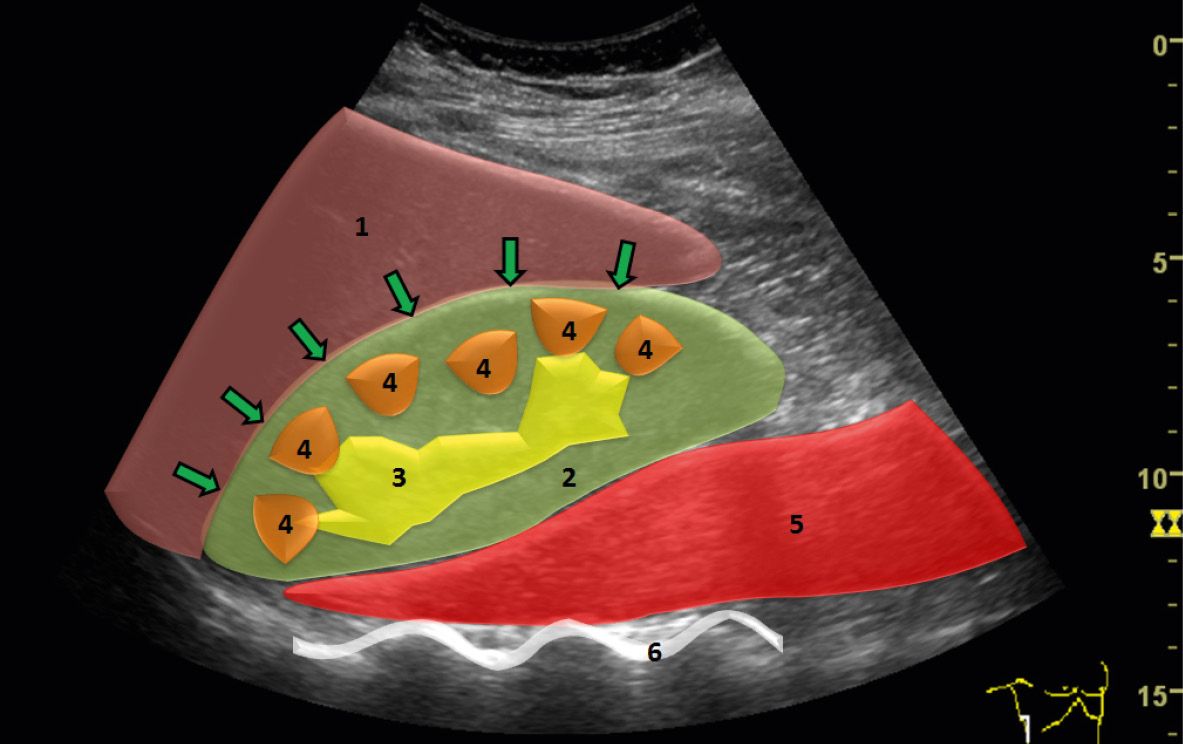
- Kidney
- Liver
- Psoas major muscle
- Spine
This section is ideal for starting an investigation and getting to know the functions of the device. The anatomical structures are usually seen clearly. One is able to obtain an overview as well as inspect smaller details when taking a closer look.
Ideal section for a beginner
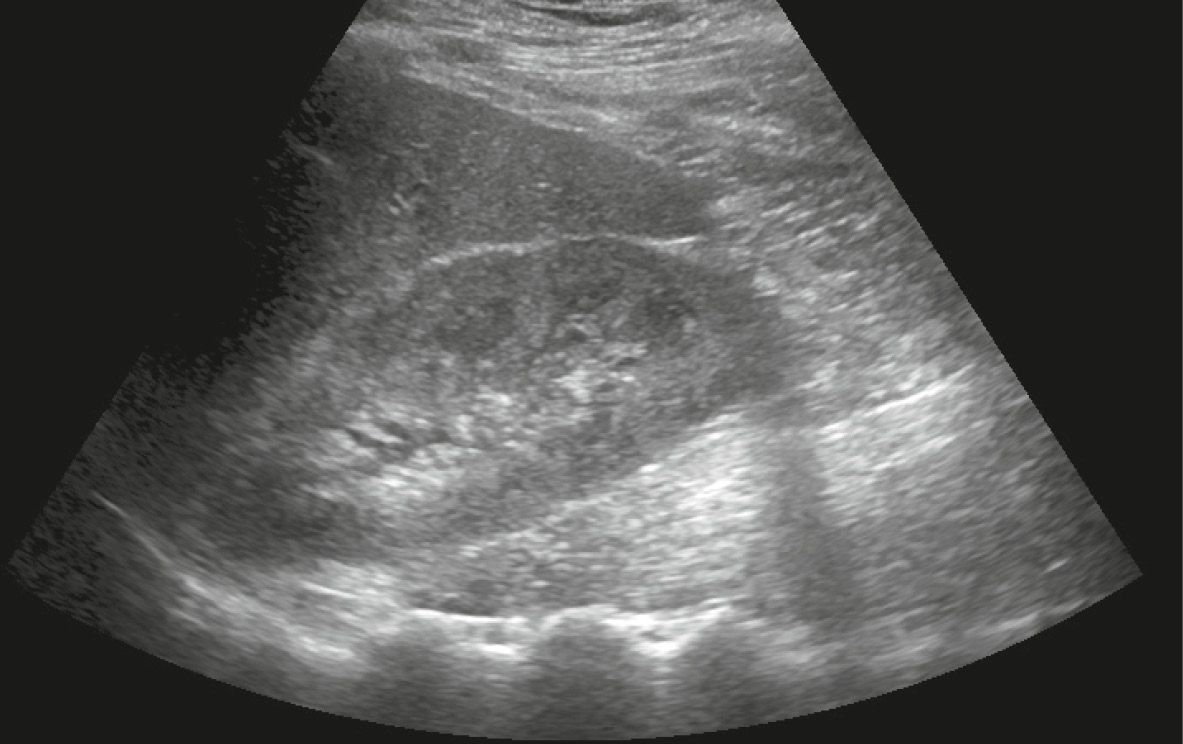
Fig. 2: Ultrasound investigation of the right flank, longitudinal section, B-mode image.
On the longitudinal section the kidney is seen centrally in the right flank. The liver is directly adjacent to it so one can compare the parenchyma of the two organs. If there is a space filled with fluid between the liver and the kidney on this section (Morison’s pouch), it is indicative of ascites or blood due to traumatic injury.
The psoas major muscle is clearly seen on the overview, as are the hyperechoic spine and its acoustic shadows. Therefore, the longitudinal ultrasound section is very suitable to get acquainted with hyperechoic bone and its dorsal acoustic shadow, as well as the striated fibers of muscle tissue.