Anomalies of Cerebral Vasculature: Diagnostic and Endovascular Considerations


Anomalies of Cerebral Vasculature: Diagnostic and Endovascular Considerations


PHILIP M. MEYERS

VAN V. HALBACH

A. JAMES BARKOVICH


INTRODUCTION

Cerebrovascular disease is uncommon in children and adolescents. In children under the age of 15, the average annual incidence of cerebral vascular disease unrelated to trauma or infection ranges from 2.5 to 3.1 per 100,000 population (1,2,3,4). Pediatric vascular disease can be grossly divided into occlusive vascular disease and causes of intracranial hemorrhage. Occlusive vascular disease and its imaging manifestations were discussed in Chapter 4. This chapter discusses cerebral vascular anomalies and cerebral vascular disorders amenable to endovascular intervention.

Vascular malformations are the cause of nearly all nontraumatic intracranial hemorrhages in children beyond the neonatal stage. Tumors, the next most common cause of hemorrhage, occur far less frequently (5,6). Therefore, any child presenting with spontaneous intracranial hemorrhage should be evaluated for child abuse (see Chapter 4) and for vascular malformations.

The treatment of pediatric cerebrovascular disease continues to advance rapidly. Endovascular techniques now permit palliation or cure of many disease entities. In vein of Galen malformations, for example, morbidity and mortality have been significantly reduced using endovascular techniques compared with conventional surgery. Consequently, a premium is placed upon the rapid recognition of treatable illness to facilitate prompt and appropriate intervention. This chapter discusses pediatric cerebrovascular disorders, with consideration toward endovascular management when indicated.


TECHNICAL CONSIDERATIONS IN PEDIATRIC NEUROANGIOGRAPHY AND INTERVENTION

Although computed tomographic angiography (CTA) and magnetic resonance angiography (MRA) can answer many questions about the vasculature of the brain, head, and neck, catheter angiography remains essential to careful analysis of disorders of the cerebral vasculature. In experienced centers with dedicated neuroangiographers, cerebral angiography is a very safe procedure, with low morbidity (7,8). Modern fluoroscopic equipment permits high-resolution biplane digital subtraction arteriography. High spatial resolution, high-speed real-time image acquisition, and high-resolution road-mapping facilitate surgical
planning and immediate treatment of complex cerebrovascular lesions. A significant level of arteriographic detail can be lost, however, due to relatively minor patient motion. Most children cannot adequately cooperate for cerebral arteriographic procedures; thus, deep sedation or neuroleptic anesthesia is an essential component of pediatric neuroangiography. While the decision to use conscious sedation or general anesthesia must be made on an individual basis, we prefer the use of general anesthesia in most cases, especially in preadolescent children. The sedation/anesthesia should be performed by an anesthesiologist with extensive pediatric experience and equipped with a complete set of pediatric cardiopulmonary life support and monitoring equipment. Monitored intravenous sedation may be substituted for general anesthesia during diagnostic angiography in the most cooperative patients. An exception to the use of anesthesia is the Wada activation test, which must be performed without sedation (9).


Preprocedural Planning

Angiography is an invasive procedure; thus, preoperative planning, as for any surgical procedure, is critical. The indications must be reviewed in light of all previous imaging studies, prior surgical procedures, and evolution of the anatomic pathology. Discussion among the angiographer, referring clinician, consultants, the patient, and the patient’s family, should result in consensus on goals and expectations for treatment. It is important that the angiographer obtain a tailored history and physical examination in order to identify specialty-specific factors such as allergies, medications, and renal or hematologic diseases that could delay or alter performance of the angiogram. Preprocedural physical examination including neurologic evaluation is also important so that any changes during the procedure can be quickly recognized and expeditiously treated. Evaluation of the extremities, including lower extremity pulses, is relevant to selection of a site for vascular access. Unique risks to pediatric angiography are the development of leg length discrepancy and claudication due to injury of the femoral artery, and femoral capital necrosis due to injury of the foveolar artery. Other complications of diagnostic angiography include stroke, iodinated contrast reaction, renal failure, and puncture site hematoma (13,14,15,16,17,18,19). In general, catheter cerebral arteriography can be performed with a minimum of risk (0.3%) at experienced centers (8).

Prior to embolization of an arteriovenous malformation (AVM), an echocardiogram possibly including a “bubble test” is warranted to evaluate for the presence of right-to-left intracardiac shunts. It is possible for embolic materials or thrombus to pass through an AVM into the normal venous drainage and the right side of the heart. Ordinarily, these small emboli will be filtered by the pulmonary vascular bed and in small numbers without any clinically significant consequences. Intracardiac shunts, common in pediatric patients with certain cerebrovascular malformations, could cause such emboli to pass from the right heart into the left heart and recirculate into the arterial system. The presence of intracardiac shunts means that great care must be undertaken during the embolization procedure to avoid the use of agents likely to pass through the cerebral AVM with resultant embolization to the systemic circulation (20).

The importance of preoperative preparation in regard to the pediatric patient cannot be overemphasized (21,22). Medications (including iodinated contrast) can cause dramatic shifts in intravascular fluid volume and vasomotor tone. An ill child may appear hemodynamically stable but, in actuality, may be exerting maximal physiological mechanisms to maintain blood pressure and systemic and cerebral perfusion. Administration of sedatives, anesthetics, or vascular contrast can cause hemodynamic collapse in a volume-depleted child. As children undergoing cerebral arteriography may have elevated intracranial pressure (ICP), adequate ventilation is essential to prevent buildup of CO2 (and consequent elevation of ICP) that could result in herniation syndromes or hinder cerebral perfusion. Occasionally, manipulation of ventilation rate and blood pressure may be necessary to modulate arterial carbon dioxide levels and vasomotor tone. Changes in the pCO2 can be used to enhance cerebral arteriographic resolution, to reduce the volume of contrast, or to assist distal microcatheter manipulation (23). However, in children with suspected ischemic stroke, especially in the setting of moyamoya disease, blood pressure should be supported while maintaining normal pCO2. These children will develop stroke if overventilated. Therefore, the pCO2 should be manipulated only by highly experienced personnel and in the presence of ICP monitoring in order to prevent cerebral hypoperfusion and herniation syndromes. Careful monitoring during the administration of sedative medications and vascular contrast is essential.


Fluid and Contrast Limitations

To limit osmotic fluid shift and to reduce the incidence of contrastinduced renal injury, contrast dose (300 mg iodine/mL nonionic contrast) should not exceed 6 mL/kg. For routine digital subtraction angiography, contrast can be diluted by 50% (to 150 mg iodine/mL) if modern fluoroscopic equipment is used. Modern “biplane” neuroangiography equipment allows acquisition of arteriographic series in two projections simultaneously. However, evaluation of high-flow arteriovenous shunts may require use of full-strength contrast. Each catheter, including access sheaths, must be continuously flushed with heparinized saline (1000 units heparin/L) through a flow-regulated system with at least 3 mL/h. However, overly exuberant flushing can cause fluid overload and congestive heart failure (CHF) in small children or children with renal impairment. The anesthesiologist should be informed of the total fluid infusion volume and rate of fluid administration through all catheters.


Vascular Access

Arterial access is the first critical step in performance of any pediatric neuroangiographic procedure. A child’s vessels are small in caliber, prone to spasm, highly mobile, and elastic. These factors often conspire to make arterial access a daunting challenge. Improper technique can result in laceration or thrombosis of the vessel. Arterial cannulation is facilitated by extension of the legs and elevation of the pelvis to straighten the common femoral artery. Following induction of general anesthesia, systemic blood pressure is often reduced, and manual palpation of the femoral and pedal pulses may become difficult. Marking the skin over the femoral puncture site as well as the foot over the dorsalis pedis and posterior tibialis pulses prior to anesthetic induction can facilitate cannulation and postoperative patient assessment. Proper positioning and padding of pressure points is necessary, particularly in infants and small children. Use of a micropuncture set and a 3 or 4 French access sheath-catheter system will help to reduce local trauma at the puncture site. Systemic heparinization (70 units/kg) during catheterization may also reduce the risk of thrombosis. Assessment of anticoagulation status in the angiography suite is rapidly performed using the activated clotting time (ACT). Depending upon the indication for angiography and other mitigating factors, an ACT 2 to 3× a baseline value is considered therapeutic. In the presence of a recent cerebral hemorrhage or surgical procedure, anticoagulation may be contraindicated. A lower level of anticoagulation for interventional procedures should be considered, if necessary. Following removal of the femoral sheath, access for subsequent arteriographic procedures should alternate between the right and left femoral arteries. In special circumstances, such as neonatal arteriography, the umbilical artery and vein readily provide vascular access for catheter systems sized up to 5 French. If so requested, the neonatology service will maintain umbilical catheters to permit arterial and venous access for endovascular procedures.


TABLE 12-1 Agents for Cerebral Embolization


Agent


Physical state


Manufacturer


Application


Size


Permanence


Polyvinyl alcohol


Particulate


Cordis, Target


Preoperative tumor, AVM embolization


45-1000 µm, calibrated sizes


++


Gelfoam sponge


Pledget (hand cut)


Upjohn


Traumatic hemorrhage


1-4 mm (cut from wafer)


+


Gelfoam powder


Particulate


Upjohn


Preoperative tumor, AVM embolization


40-60 μm


++


Avitene


Particulate


Medchem


Preoperative tumor, AVM embolization


75-150 μm


+


Embospheres


Particulate


Biosphere Medical


Tumor embolization


40-1200 μm, calibrated sizes


+++


Platinum coils


Metallic coil


Cook, Target


Tumor, AVM


Multiple


++++


Detachable platinum coils


Metallic coil


Target


Aneurysm, AVM


Multiple


++++


Balloons


Latex


Balt


Fistula


Multiple


+++


Cyanoacrylate


Liquid adhesive


J&J, Codman


AVM, Fistula


N/A


++++


Ethylene vinyl copolymer


Liquid precipitate


Ev3


AVM, Fistula


N/A


+++


Ethanol


Sclerosant


Abbott


Fistula, AVM


N/A


++++


Ethanolamine oleate


Sclerosant


Questcor


Hemangioma, venous, and lymphatic malformations


N/A


++++


Catheter Choice

The choice of angiographic catheter depends upon the application and patient size. The smallest diameter catheter that readily permits diagnostic arteriography is a 3 French catheter, which is used in neonates and infants. Smaller diameter catheters do not generally allow sufficient injection rate for routine arteriography. Slightly larger (4 French) catheters can be used in older children and provide greatly improved injection rates due to decreased flow resistance. Standard microcatheters can be advanced either through a 4 French diagnostic catheter platform in the cervical vessels or directly through a 4 French access sheath in the femoral artery without coaxial passage into the craniocervical region. Older children and adolescents will tolerate the use of the 4 to 6 French catheter systems commonly used in the adult population. Percutaneous access sheaths are used routinely in our practice to facilitate catheter exchange and to reduce trauma at the puncture site.


Endovascular Occlusion

The purpose of interventional neuroradiology is the minimally invasive treatment of vascular lesions. The fundamental principle of this treatment is occlusion of pathological vascular channels to improve the patient’s neurological or cardiovascular condition. Embolization, or endovascular occlusion, requires placement of the tip of a microcatheter in proximity to the abnormal vessels, so that these channels can be selectively occluded, sparing normal or other needed vessels. Recent and on-going developments in catheter technology allow careful and precise placement of embolic materials within the abnormal vascular channels while avoiding embolization of the normal adjacent vessels. A number of embolic materials are currently in use and many more are under development or employed in clinical practice outside the United States. Table 12-1 represents a listing and some characteristics of some
of the more commonly used embolic materials. A complete review of the specific applications of these embolic agents is beyond the scope of this text.


Postprocedural Patient Care

At completion of an arteriographic procedure, the femoral access catheter must be removed and the puncture site must be manually compressed. Arterial closure devices generally require an artery that is at least 5 to 6 mm in diameter to be deployed safely. Ideally, the catheter is removed while the patient is still under anesthesia to assure optimal control of the puncture site. Continuous Doppler monitoring of the pedal pulses is recommended to prevent excessive pressure with inadvertent thrombosis of the femoral artery during manual compression. Hemostasis is usually attained within 15 minutes in a patient with normal coagulation parameters. An anticoagulated patient may require the administration of protamine sulfate (10 mg protamine per 1000 units of active heparin in the blood) to reverse the effects of heparin prior to removal of the access catheter. Coagulopathy for other reasons may require administration of blood products or synthesized hemostatic factors. With an ACT below 180 seconds, it is usually possible to obtain arterial hemostasis. Thereafter, the patient should be closely monitored for several hours for evidence of puncture site bleeding, subcutaneous hematoma, or loss of distal pulses.


INTRACEREBRAL HEMORRHAGE IN CHILDREN

In contrast to adults, intraparenchymal hemorrhage is at least as common as ischemic stroke in children and it is an important cause of morbidity and mortality (24,25,26). Trauma is the most common cause of intraparenchymal hemorrhage; the cause can be deduced from history or external evidence in these patients. Vascular malformations seem to be the most common cause of spontaneous cerebral hemorrhage in children, with true AVMs accounting for about 25%, and cavernous malformations about 20% (27). Tumors account for about 10% (usually ependymoma or high-grade glioma or PNET) and venous sinus thrombosis for another 10% (27,28); venous thrombosis seems more common in infants and neonates (28,29,30). Aneurysms are an uncommon cause, accounting for less than 5% (27,31). Most of the remaining cases are the result of systemic disease, such as infection, leukemia, or severe thrombocytopenia (27,32).

CT remains the primary imaging modality in the initial diagnosis of intracranial hemorrhage. After the presence of hemorrhage has been established, MRI with MRA and MRV can often establish the cause of hemorrhage. MRI with and without contrast may reveal enhancing tumor, if present, and may show anatomically remote cavernomas or an adjacent developmental venous anomaly that will allow a diagnosis of cavernous malformation. Large aneurysms or vascular malformations will also be evident by MR or MRA. Venous thrombosis can be detected by hyperintense signal in the vein on T1-weighted images or on MRV (more discussion of venous thrombosis is found in Chapters 4 and 11). If the MRI is unrevealing, diagnostic catheter angiography becomes an important tool to look for small vascular malformations (33,34).


INTRACEREBRAL VASCULAR MALFORMATIONS

Intracerebral vascular malformations (AVMs) are malformations of the cerebral vasculature that may involve arteries, capillaries, or veins. Although a few reports of familial vascular malformations have been reported (35), the majority are sporadic. Pathologically, vascular malformations have been subdivided into four groups. In McCormick’s series of 248 children with vascular malformations, AVMs were found in 12%, developmental venous anomalies (DVAs) in 62%, cavernous malformations in 8%, and capillary telangiectasias in 18% (36). Vein of Galen malformations, a type of AVM specific to neonates and infants, and facial AVMs are discussed separately, as special cases of AVMs.


Arteriovenous Malformations


Description and Causes

AVMs are compact collections of abnormal, thin-walled vessels that connect dilated arteries to veins without an intervening capillary network. These persistent primitive connections between arteries and veins are formed during the late somite stages of the fourth embryonic week; they develop from the sinusoidal vascular network that perfuses the early developing telencephalon. The absence of capillaries produces a low-resistance shunt, which results in rapid arteriovenous shunting within the malformation.

Patients with cerebral AVMs most commonly present as a result of spontaneous intracerebral hemorrhage (up to 61% of cases) during late childhood (37) or as a young adult (38), but affected patients can present at any age or, rarely, with seizures (39). Intraparenchymal hemorrhage can cause significant injury at any age, with 30% to 50% neurological morbidity and 10% mortality (40). Consequently, a premium has been placed upon identification and treatment of AVMs at major neurosurgical centers throughout the world.

The prevalence (total number of cases in a population at a given time) of AVMs has been estimated in autopsy series between 0.06% and 0.11% (38,41,42,43). Symptomatic AVMs were detected in 1 per 100,000 person-years, and prevalence was inferred from the incidence data to be less than 10 per 100,000 in a US population study (44,45) and 5 per 100,000 in Australia (46). Thus, although the precise numbers are not known, it seems that a significant percentage of AVMs become symptomatic during the average human lifetime, usually due to hemorrhage (38,46,47).

The vascular channels of AVMs that develop instead of normal capillaries are characterized by fibrointimal thickening and elastic tissue disorganization (48). These features probably take time to develop and, thus, classic brain AVMs (those with the classic tangle of vessels that compose the nidus) are not typically seen before birth or in early infancy. No classic AVMs have been described prenatally, although three fetal cases with pial fistulas (direct connections between artery and vein without the tangle of abnormal vessels) have been identified (40). In neonates, AVMs are generally seen as direct arteriovenous communication without intervening nidus. Thus, AVMs seem to evolve, not becoming fully developed until later in life (49). No definite genetic linkage has been identified with the exception of Rendu-Osler-Weber (50) and Wyburn Mason syndromes (51). Birth-related or other trauma has been suggested as an inciting event (52), but this is not supported by experimental data. Dysautoregulation of vascular growth factors has been implicated in the subsequent development and maturation of the vascular malformation (53). De novo development of AVMs has rarely been documented in children or adults (54,55).

AVMs can increase in size with age, as a result of increase in size of the nidus and progressive dilation of the feeding arteries and draining veins. Astrogliosis and atrophy is often seen in surrounding brain tissues. This was at one time thought to be a consequence of low-resistance shunting of blood through the nidus, causing parenchymal hypoperfusion (vascular steal). However, current thought suggests that edema and subsequent parenchymal injury results from venous stenosis/venous
hypertension, caused by high pressure and shear stresses from turbulent blood flow and consequent stenosis in draining veins (56,57). Thus, contrary to earlier descriptions, AVMs can have mass effect unassociated with hemorrhage. Rapid flow in the feeding arteries may result in the development of arterial aneurysms, both in the same distribution as classic berry aneurysms (circle of Willis) and in feeding arteries (pedicle and nidal aneurysms) close to the malformation (Fig. 12-1). In addition, varices (venous aneurysms) can develop upstream to the stenosis and cause hemorrhage if they rupture. As a result of all these factors, AVMs are the vascular malformations that most commonly cause neurological defects other than seizures, despite the fact that they are not the most common form of vascular malformations.


Clinical Manifestations

Patients with AVMs usually present with seizures, recurrent headaches, progressive neurological deficits, hydrocephalus, or hemorrhage. Approximately 20% become symptomatic before the age of 20 years (37). The mortality associated with the initial rupture of an AVM is 10%, with morbidity between 30% and 50% (36,58). Morbidity and mortality increase with each subsequent hemorrhage.


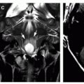
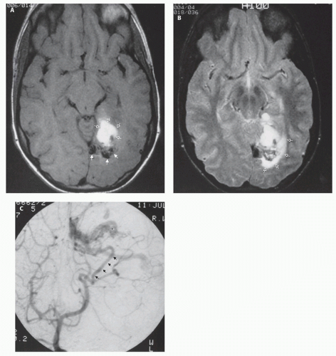
FIG. 12-1. AVM with hemorrhage. A. Axial T1-weighted image shows an area of high signal intensity in the left temporooccipital region (open arrows). This subacute hematoma lies directly ventral to a tangle of vessels (solid arrows), which presumably represents the nidus of the AVM. B. Axial T2-weighted image shows the tangle of vessels and hematoma. The area of high signal intensity (open arrows) dorsal and lateral to the tangle of vessels represents a small amount of edema. C. AP image from a left vertebral artery angiogram shows the vascular malformation being fed by the left posterior temporal artery (solid arrows).

AVMs are the cause of up to 40% of spontaneous intracranial hemorrhages (2,59,60), most of which occur in the cerebral parenchyma. However, superficial malformations can rupture into the subarachnoid space and deep malformations may cause intraventricular hemorrhage. Symptomatic vasospasm and rebleeding are relatively rare unless the source of hemorrhage is an associated aneurysm. The risk of rehemorrhage from an AVM has been estimated to be higher in children than adults (58). In children less than 15 years of age, AVMs are the most common cause of spontaneous intracranial hemorrhage and account for 20% of all strokes (2).

Seizures occur in approximately 70% of patients with AVMs, with one half of the seizures being generalized (61); most patients are well controlled with anticonvulsants. Seizures are most common in AVMs that are located in the cerebral cortex and have associated varices. Their
cause is probably cortical injury and gliosis from previous hemorrhage (62) or venous ischemia (63,64).

Chronic recurrent headaches may be the presenting symptoms in some patients with AVMs, and are likely caused by engorged dural vessels. Headaches are most common in cortical/subcortical AVMs and in those that develop stenosis or occlusion of feeding cerebral vessels. AVMs located in the occipital region are associated with an increased frequency of migraines, often with visual symptoms during the migrainous episodes.

Progressive neurological deficits occur in a small percentage of children with AVMs, particularly if the malformation is large and located close to the motor cortex. The probable cause of neurologic deficit is venous hypertension, as increased venous pressure is transmitted through the AVM, impairing the venous drainage of normal brain sharing the same venous drainage pathway (65). This mechanism has long been understood as a cause for neurologic deficit in the spinal cord, but has only recently been recognized in the brain. Venous hypertension may be so marked that it impairs the resorption of cerebrospinal fluid, producing hydrocephalus (see Chapter 8).

Finally, with the advent of CT and MRI, asymptomatic AVMs are more commonly discovered.


Imaging Findings

The diagnostic workup of vascular malformations usually includes x-ray CT, MRI, and MRA. CT evaluation (using a low dose, see Chapter 1) remains the standard of care as an effective screening technique to study patients with suspected intracranial hemorrhage in the acute phase (<2 weeks). A noncontrast CT scan may reveal abnormal high density of the associated acute hematoma or calcifications within the interstices of the AVM. In addition, CT may show acute subarachnoid hemorrhage, which sometimes accompanies rupture of a superficial AVM or an associated aneurysm. During the ensuing 1 to 2 weeks after hemorrhage, the subacute hematoma becomes progressively isointense with brain and may show a rim of peripheral enhancement following intravenous contrast administration; this appearance is nonspecific and may be mistaken for infection, cavernoma, or neoplasm. While MRI has largely supplanted CT in the evaluation of AVMs, vascular malformations may be detected on contrast-enhanced CT as a region of enhancement adjacent to a high-density, acute hematoma. CTA shows detailed resolution of the cerebral vasculature, but can be associated with significant radiation exposure. Its role in the architectural evaluation of AVMs remains undefined.

MRI is an excellent initial screening modality for the evaluation of intracranial AVMs (66,67). Fast-flowing intraluminal blood in AVMs appears as regions of decreased signal or “flow void” on spin-echo sequences. A tangle of serpiginous flow-void represents the nidus of the malformation (Fig. 12-1). Rapid flow within draining veins can be difficult to distinguish from feeding arteries in or around the nidus. The multiplanar capability of MRI helps to delineate the location of the malformation in relation to critical anatomic structures. Parenchymal hematomas evolve through a recognizable and often specific pattern of signal characteristics (see Chapter 4 [68,69]). The residua of previous hemorrhages adjacent to malformations remain detectable many years after hemorrhage as areas of very low intensity on T2 or T2*-weighted images. In the subacute and chronic phases of intraparenchymal hemorrhage, MRI is more sensitive and specific than CT for the detection of blood products. In patients with AVMs, rapid sequence catheter angiography must still be performed to delineate the angioarchitecture, distinguish feeding arteries from draining veins, and to identify any associated arterial aneurysms or venous restriction that would be associated with a poorer natural history.


Treatment

The decision to undertake treatment for symptomatic AVMs should consider the natural history of the disease and the projected risks and benefits of the therapy. A complete discussion of the natural history and current therapeutic options is beyond the scope of this book, and the reader should consult a more complete text for the discussion of management (70,71,72). Briefly, the therapeutic options fall into three categories: surgical excision, radiosurgery, and endovascular embolization or occlusion. Often, combinations of these techniques are utilized for the treatment of complex malformations.

With the improvement in microneurosurgical techniques, even large and complex malformations can be excised with relatively low morbidity and mortality (70,73). The nidus or core of the malformation infrequently contains cerebral parenchyma and, if the surgical excision is performed along the margins of the malformation with attention to preserving the surrounding brain, an excellent outcome can often be achieved. Total excision of the malformation is the goal of surgery, as prior studies have shown that subtotal resection or ligation of feeding vessels is inadequate to prevent subsequent hemorrhage (74,75).

Radiation therapy produces hyperplasia of the blood vessel walls; the AVM is completely obliterated in selected cases. To minimize the risk of radiation-induced damage to adjacent normal parenchyma, only focused techniques are utilized today, such as the Bragg-peak proton beam therapy or intersecting beam techniques using gamma knife or LINAC (linear accelerator) therapy units. Occlusion rates range from 35% to 92% depending on a number of variables including AVM size, location, and radiation dose. The results of radiotherapy have been quite encouraging with small (<3 cm) malformations (76,77,78,79,80). The major disadvantages of radiosurgical techniques include the difficulty in treating larger lesions, the 1 to 2 year latency before development of significant vascular intimal hyperplasia and fibrosis (during which interval the patient remains at risk for hemorrhage), and the risk of delayed radiation necrosis in the surrounding brain tissue.

Endovascular techniques have emerged as a third modality for the treatment of symptomatic intracranial malformations (81). Complete angiographic obliteration following embolization of large malformations is unusual; however, complete obliteration can be obtained with smaller AVMs (82). Endovascular embolization can palliate patients whose symptoms do not warrant more aggressive therapy or where the size or location of the malformation precludes more traditional forms of therapy. These procedures can now be performed with a low risk of complications and appear to make craniotomy for surgical resection safer (70,83). Patients who present with intractable unilateral headache frequently have recruitment of dural vascular supply to the malformation (82,84). These dural vessels can be selectively catheterized and embolized, often resulting in alleviation or resolution of headache. Patients with progressive neurological decline from arterial steal or venous hypertension can demonstrate clinical improvement following partial embolization of their malformation.

The vast majority of AVM embolization procedures performed at our institution are performed as a preoperative adjunct (70). The most commonly used embolization agent is the liquid acrylic, n-butyl cyanoacrylate. A relatively new embolic agent preferred by some neurointerventionalists called Onyx (eV3, Maple Grove, MN) is a precipitate of ethylene vinyl alcohol suspended in an organic solvent (DMSO). While high rates of nidal occlusion are possible with this agent, complication rates appear comparatively higher based on currently published series (85). By occluding the deep or inaccessible feeders to the malformation, surgical excision is facilitated (86). Preoperative embolization can reduce the size and pressure within the nidus, making subsequent surgical excision easier and reducing intraoperative blood loss (86).
In larger malformations, the adjacent brain parenchyma can lose its ability to autoregulate; thus, staged palliative embolization is used to gradually re-establish regulatory control in the microcirculation and prevent the development of normal perfusion pressure breakthrough (86). In reducing the size and rate of flow through a malformation, improved obliteration of AVMs may be achieved by radiosurgery, although posttreatment evaluation is warranted.

We and others (87,88) have identified a group of patients with solitary arteriovenous connections who present in childhood with hemorrhage or neurologic deficit (Figs. 12-2 and 12-3). All 15 patients were treated by transvascular embolization techniques with placement of either platinum coils or balloons at the fistula site. Fourteen of the fifteen patients were completely cured and the fifteenth patient underwent surgical excision without incident.


Vein of Galen Malformations


Description and Causes

Malformations involving the vein of Galen are rare congenital connections occurring between intracranial arteries (usually thalamoperforator, choroidal, and anterior cerebral arteries) and the vein of Galen or other primitive midline vein (37,69). These connections can be large direct fistulas, numerous small connections, or a combination thereof. The cause of these connections is unknown. Some investigators have noted a strong association with venous anomalies (absent straight sinus, persistent falcine and occipital sinuses) and suggested that intrauterine straight sinus thrombosis with recanalization is responsible (69). Raybaud et al. demonstrated that the dilated primitive venous structure represents persistence of the embryonic median prosencephalic vein of Markowski (89). They suggested that early obstruction of the straight sinus might result in persistence of the primitive veins based upon the need for venous drainage pathways. Moreover, in our experience, vein of Galen malformations are associated with certain cardiovascular anomalies, most commonly aortic coarctation and secundum atrial septal defects (90).


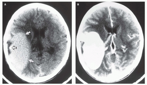
FIG. 12-2. A solitary arteriovenous connection (fistula). A. Axial noncontrast CT scan shows a large mass in the right sylvian area (solid arrows) that is hyperdense compared with brain parenchyma. A small focus of calcification (open arrow) is present within the mass. B. After infusion of iodinated contrast, the mass uniformly enhances. C. Coronal T1-weighted image shows a large amount of signal misregistration (arrows) from the mass in the phase-encoding direction. This phase misregistration artifact is essentially pathognomonic for a vascular lesion. D. Arterial phase image from a right internal carotid arteriogram, lateral projection, shows an enlarged Rolandic branch of the middle cerebral artery emptying into a large venous varix at the site of the solitary A-V connection.

Over 90% of the vein of Galen varices falls into the group called “choroidal” malformations (91). Choroidal malformations are arteriovenous connections from a plethora of vessels, usually numerous choroidal, pericallosal, and thalamoperforator vessels, to the anterior wall of the prosencephalic vein, resulting in a great deal of arteriovenous shunting (92,93); the consequence of the shunting is presentation as neonates with CHF. Choroidal malformations have the poorest prognosis, and are usually fatal without treatment. The second, less common, category of vein of Galen malformation is the so-called “mural” malformation (91). Mural malformations are characterized by fewer (usually one to four) but larger caliber connections with the prosencephalic vein; the posterior choroidal or collicular arteries are most commonly involved. Patients with mural malformations usually present in infancy with developmental delay, hydrocephalus, and seizures but mild or no signs of CHF. The treatment approach to these malformations varies although endovascular therapy has become the method of choice for both and offers a high rate of cure with low morbidity (94,95,96).


Clinical Presentation and Imaging Findings

The clinical presentation of vein of Galen malformations can be categorized into three groups: (i) the neonate presenting with intractable CHF and loud intracranial bruit, (ii) the infant presenting with hydrocephalus and/or seizures, and (iii) the older child or young adult presenting with hemorrhage (97). As mentioned in the preceding section, patients in group 1 typically have choroidal malformations, whereas patients in groups 2 and 3 have mural malformations.


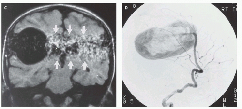
FIG. 12-2. (Continued)

With improvements in quality and availability of prenatal imaging, many vein of Galen malformations are now diagnosed antenatally (98,99). Prenatal sonograms show a large hypoechogenic to mildly echogenic midline mass that is seen to have rapid flow on Doppler studies. MRI identifies the varix best (Fig. 12-4) and feeding vessels, and can be used to identify any associated structural abnormalities such as malformation or ischemic injury. If such a patient is identified, the interventional neuroradiology service must be alerted and available to provide assistance at the time of delivery and in the perinatal period. If the patient is found to have neonatal heart failure that is refractory to medical therapy, the neurointerventionalist may need to treat these patients (100).


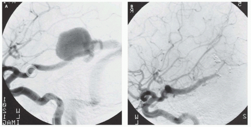
FIG. 12-3. A fourteen-year-old girl with severe unilateral headaches. A. Left internal carotid angiogram, lateral projection, shows a solitary fistula arising from the posterior temporal branch of the posterior cerebral artery. B. Same injection and projection, status post coil embolization, confirms complete closure of the fistula.

If the vein of Galen malformation is not diagnosed prenatally, postnatal neuroimaging becomes critical to making the proper diagnosis. On imaging studies, the dominant feature of the vein of Galen malformation is the varix, which appears as a large mass in the incisural region, sometimes extending rostrally and anteriorly displacing the third ventricle (Fig. 12-4). On sonography, the varix will appear mildly echogenic on two-dimensional (2D) imaging and show turbulent flow on Doppler (Fig. 12-5); it is important to demonstrate continuity with the straight sinus or a persistent falcine sinus. Doppler studies may be useful to quantify the rate of flow within the varix for future reference. On CT, the varix will be iso- to hyperdense to brain prior to contrast administration (Fig. 12-5). Mixed attenuation may be seen if the varix is partially thrombosed. Areas of low attenuation (encephalomalacia, usually secondary to ischemia) and high attenuation (hemorrhage or dystrophic calcification) are often present in the brain parenchyma. The extent of brain injury should be carefully analyzed, and the parents informed of likely neurological and developmental sequelae, before therapy is begun. On MRI, the varix will be hypointense (Fig. 12-4) resulting from a loss of phase coherence of the mobile protons; mismapped signal will often be seen across the image in the plane of the phase-encoding gradient (Figs. 12-4 and 12-6). Feeding vessels will be identified on axial images as round areas of signal void in the ambient cisterns and on MRA as thin, curvilinear structures demonstrating flow-related enhancement and connecting to the varix. Areas of acute thrombosis will usually be isointense to brain on T1-weighted sequences and hypointense on T2-weighted sequences, whereas subacute thrombus will have a high intensity on both T1- and T2-weighted sequences. Thrombus of varying age usually lines the wall of the varix. Areas of damaged brain will typically appear hyperintense on T1-weighted images and hypointense on T2-weighted images in the neonate (Fig. 12-6); they may be difficult to see on FLAIR. Diffusionweighted images will show acute injury as regions of reduced diffusivity (Fig. 12-6). As the brain begins to myelinate, injured brain is better seen on T2-weighted images and then on FLAIR images (see explanations in Chapter 4). If left untreated, the fistula will continue to grow, recruiting additional blood supply and engendering new fistulas (Fig. 12-7).


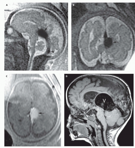
FIG. 12-4. Fetal and postnatal MRIs of vein of Galen malformations. A-C. Fetus of 29 week gestational age. Sagittal T2-weighted image (A) shows a large varix (black arrow) above the quadrigeminal plate cistern. The straight sinus (black arrowhead) appears normal or enlarged. Coronal T2 (B) and axial T1 (C) images show the large varix (black arrows). It appears hyperintense in (C) because of inflow of unsaturated protons on this spoiled gradient-echo image. E-H. A two-year-old boy with increased head circumference and Parinaud syndrome (paralysis of upward gaze due to compression of the brainstem tectum). D and E. Sagittal T1-weighted and axial T2-weighted images reveal marked enlargement of a central venous structure (the dilated median prosencephalic vein of Markowski, V) with hydrocephalus resulting from compression of the dorsal midbrain and cerebral aqueduct. F. Left vertebral artery injection, frontal projection in the arterial phase, demonstrates a mural-type vein of Galen malformation supplied by the collicular arteries through a single-hole fistula (curved arrow). G. Left vertebral injection, lateral projection in the late venous phase, shows to advantage the dilated prosencephalic vein of Markowski draining into a persistent falcine sinus. In these cases, the straight sinus is often absent, and it has been proposed that intrauterine thrombosis of the straight sinus results in the vein of Galen malformation. H. Left vertebral injection, frontal projection in the arterial phase following occlusion of the right and left collicular arteries, show complete occlusion of the malformation. The patient made an uneventful recovery and is now neurologically normal.


FIG. 12-4. (Continued)


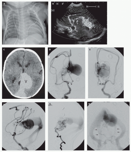
FIG. 12-5. Newborn with severe intractable congestive failure. A. Portable chest radiography demonstrates cardiomegaly and increased pulmonary vascular markings compatible with pulmonary edema due to high-output CHF apparent on physical examination. B. Transfontanelle color Doppler ultrasonography, sagittal plane, shows prominent anterior cerebral arteries in continuity with an enlarged deep central venous structure. C. Nonenhanced CT brain scan confirms the presence of markedly enlarged central venous structures (V), normal brain development, and the absence of hydrocephalus or hemorrhage. D-H. Complete cerebral arteriography in multiple projections demonstrates a choroidal-type vein of Galen aneurysmal malformation supplied by every major vascular distribution. Arteriovenous shunting converges upon a dilated primitive prosencephalic vein of Markowski continuous with a persistent falcine sinus. Presumably, due to the high-flow state, venous drainage includes persistent occipital sinuses (arrows, H).


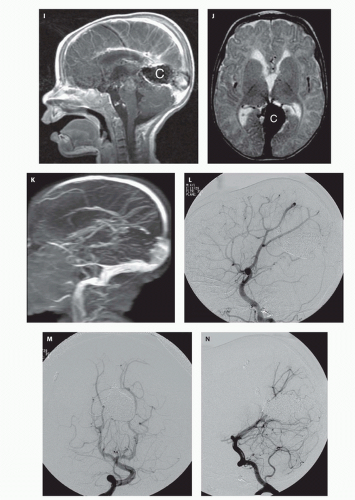
FIG. 12-5. (Continued) I-K. Following curative endovascular occlusion of the malformation, MRI brain scan demonstrates the coil mass (C), normal flow through the mature cerebral venous drainage system, the absence of cerebral infarction, and no hydrocephalus. Maximum intensity projection from 2D time-of-flight MR venogram shows intact superficial venous drainage. Drainage of the deep venous system is obscured by susceptibility artifact from the coils. L-N. Complete cerebral arteriography following both transarterial and transvenous embolization confirms complete absence of arteriovenous shunting and durable occlusion of this malformation. The child is now 4 years old, has a normal neurological exam, and meets all developmental milestones. Endovascular techniques are now the preferred treatment method for these lesions.


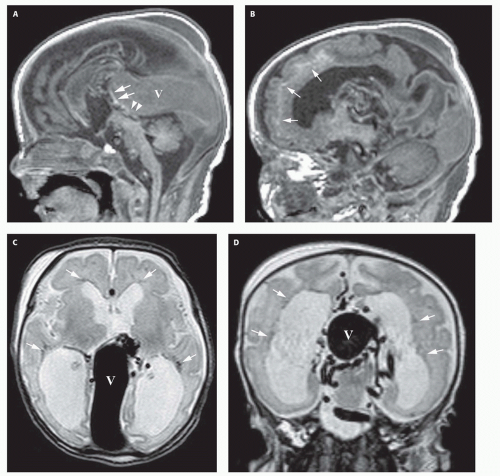
FIG. 12-6. Damaged brain in a vein of Galen malformation. This newborn presented with CHF. A. Sagittal T1-weighted image shows the large curvilinear varix (V) that compresses the aqueduct (white arrowheads), resulting in hydrocephalus, and anteriorly displaces the posterior wall of the third ventricle (white arrows). B. Parasagittal T1-weighted image shows abnormal subcortical hyperintensity (white arrows), indicating parenchymal injury. C and D. Axial (C) and coronal (D) T2-weighted images show the varix (V), multiple enlarged tortuous vessels around the midbrain and the varix, and abnormal hypointensity of the periventricular white matter (white arrows), indicating parenchymal injury/ necrosis. In addition, the ventricles are enlarged, likely secondary to a combination of hydrocephalus and loss of periventricular white matter volume. E. Average diffusivity (Dav) image shows reduced diffusivity (low signal intensity, arrows) in the posterior frontal periventricular and deep white matter. F and G. Partition image from time-of-flight MR angiogram (F) and maximum intensity projection from MR venogram (G) show the mural type of vein of Galen varix with enlarged anterior cerebral artery (large white arrowhead) and multiple branches and feeders from the posterior circulation (small white arrowheads) terminating in the varix (V). Note the enlarged emissary veins (white arrows in G) helping to shunt blood from the engorged intracranial venous system into extracranial veins.


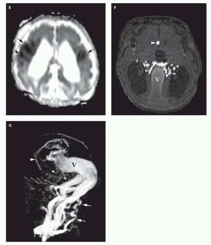
FIG. 12-6. (Continued)


Treatment

Aggressive medical management of the cardiac failure associated with Galenic malformations is an essential adjunct to surgery or endovascular procedures, but medical management alone can rarely control the failure. Johnston’s review of neonates presenting with CHF revealed a mortality of 95%, and none were stabilized without surgical intervention (101). Surgical ligation of the anomalous connections has been described, but the results have been disappointing. In a review of 60 neonates treated by surgery, there were only six survivors; half of the survivors suffered from neurological deficits (101). Several series have reported the efficacy of endovascular procedures as a palliative or definitive treatment (102,103,104). In the early 1980s, these procedures consisted of free particle embolization. While the majority of these emboli would lodge in the fistulous connections, the risk of an errant embolus occluding a normal cerebral blood vessel was inversely proportional to the flow in the fistula, and the majority of these procedures were palliative. With the development of newer microcatheter delivery systems and embolic agents such as platinum coils (105), silk sutures, and liquid adhesives (91), superselective embolization of the fistula connections alone can be achieved (Figs. 12-5 and 12-8). Mickle et al. developed a technique where the torcular is surgically exposed, a small catheter is placed transvenously through the straight or falcine sinus into the involved vein of Galen, so metal coils can be deposited to diminish the arteriovenous shunting (104). Although useful in the presence of bilateral transversesigmoid or occipital sinus hypoplasia or occlusion, the transtorcular approach is not often necessary, as embolic agents can be placed via transfemoral venous access.

Over the past 10 years, 34 children with Vein of Galen malformations have been treated at our institution; 26 harbored the choroidal variety and presented with CHF (94,103,106,107). The first five patients were treated by craniotomy and attempted clipping of the feeding
vessels. All five patients died during or shortly after the surgery. The subsequent eight patients underwent transvascular embolization techniques. Six of the eight survived while two died despite treatment (103). Of the survivors, one suffered a severe middle cerebral infarct resulting from an errant embolus. Another had a partial visual field deficit, presumably a result of ischemic damage secondary to the underlying disease. The remaining patients are neurologically and developmentally normal with marked reduction of the fistula flow following treatment. At long-term independent follow-up, 61% of patients who survived their initial presentation were neurologically normal or demonstrated only minor developmental delay (96).


FIG. 12-7. A thirty-two-year-old woman with vein of Galen malformation and presenile dementia. A and B. Midsagittal T1-weighted and axial T2-weighted images show severe calvarial thickening from chronic anticonvulsant use and extensive flow voids along the posterior falx cerebri and tentorium cerebelli, surrounding the primitive median prosencephalic vein and persistent falcine sinus. The basal ganglia hyperintensity probably represents injured, calcified tissue from chronic venous hypertension. C. 3D Phase-contrast MRA illustrates the dramatic increase in the size of intracranial blood vessels caused by the congenital vein of Galen malformation and subsequent recruitment of additional blood supply from all vascular distributions. D-F. Left internal carotid, external carotid, and vertebral injections, lateral projection in the arterial phase, define the recruitment of blood supply to the fistula from all vascular distributions. Chronic venous hypertension has engendered development of several independent and noncontiguous fistulas of the superior sagittal sinus (black arrow) and falx cerebri (white arrowhead).


FIG. 12-7. (Continued)


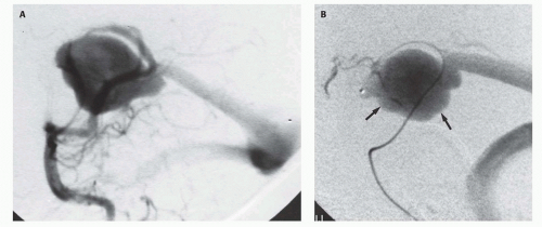
FIG. 12-8. A three-month-old infant presents with Parinaud syndrome (paralysis of upward gaze) and an enlarging head circumference. A. Left vertebral artery injection, lateral projection, demonstrates a mural type vein of Galen malformation with two large-caliber connections. B. A microcatheter has been navigated into the fistula site and contrast injected delineating the draining prosencephalic vein (arrows). C-E. Superselective injection following embolization with microcoils demonstrates complete occlusion of the fistula. Note preservation of all normal vessels, including the choroidal vessels, parietooccipital branches, and connection to the distal splenial and pericallosal artery.

Improvements in technique and embolic materials allow vein of Galen malformations to be treated by transarterial or transvenous approaches. These new materials and approaches have resulted in a high rate of successful therapy; indeed, greater than 50% angiographic cure and 75% symptomatic improvement have been achieved in our recent patients, even in those with the high-flow choroidal malformations (106,107). Our cure rate is 100% for the less common mural type of vein of Galen malformation that most commonly presents later in infancy with hydrocephalus, seizures, and failure to thrive (94).

The following are the current recommendations for treatment of vein of Galen malformations presenting with severe congestive failure.
If the diagnosis is established prenatally, the delivery should be performed at an institution offering endovascular techniques to palliate the patient should intractable CHF develop. Severe heart failure in utero can result in polyhydramnios and hydrops fetalis, which can be an indication for induced delivery. Close coordination among the obstetricians, neurointerventionalists, neonatologists, and neurosurgeons is essential to optimize planning. Baseline ultrasonography with color flow Doppler should be performed to serve as a baseline for blood flow in evaluating the results of the endovascular techniques. If possible, umbilical arterial and venous catheters should be placed at the time of delivery to allow repeated vascular access for both diagnostic and therapeutic procedures. These indwelling catheters obviate the necessity for repeated femoral punctures in the fragile neonatal femoral artery. A CT or MRI should be performed to assess any parenchymal damage already produced by the congenital fistula, disclose hydrocephalus, which may require ventriculoperitoneal shunting, and serve as a baseline.


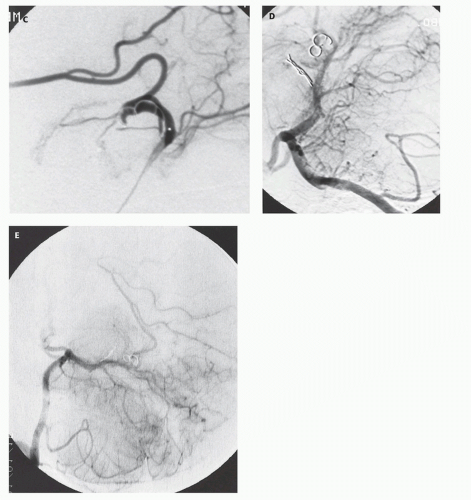
FIG. 12-8. (Continued)

If intractable congestive failure persists despite aggressive medical management, angiography is performed to delineate the vascular anatomy. Palliative arterial embolization can be performed at this time, preferably with superselective catheterization of each feeding pedicle to reduce the risk of ischemic damage to normal surrounding brain. The embolization procedure may be repeated if congestive failure persists. If the congestive failure continues and further arterial embolization is considered risky or technically impossible, transvenous embolization may become necessary. An arteriogram is performed to localize the draining venous sinuses. If the draining venous sinuses are patent, the transfemoral or transumbilical routes can be used to access the varix. On rare occasions, when the lateral sinuses are absent or severely hypoplastic, surgical access to the intracranial venous system is necessary. A small burr hole is made over the draining falcine or straight sinus. A needle puncture is made into the sinus, a catheter advanced into the varix, and platinum coils deposited. These techniques can be curative or palliative. When palliative, they alleviate the congestive failure and allow the child to develop normally until a definitive treatment can be performed with further surgical or radiological techniques. MRI scanning is useful in our experience to assess brain development, the degree of thrombosis in the fistula site, and delayed development of hydrocephalus.


Cavernous Malformations


Definition and Causes

Cavernous malformations, also called cavernomas, are spherical collections of sinusoidal (cavernous) vascular spaces (36). Multiple cavernous hemangiomas are common and are associated with a familial predisposition. Three causative genes have been identified: KRIT1 (CCM1) at chromosome 7q11.2-q21 (108); CCM2/malcavernin at chromosome 7p13 (51,109); and PDCD10 (CCM3) at chromosome 3q26.1 (51,109). The proportions of families linked to these loci have been estimated to be close to 40% (CCM1), 20% (CCM2), and 40% (CCM3) (109). The proportion of patients with onset of symptoms before the age of 15 years is highest in the CCM3 group (109), but the number of affected individuals per family is lowest in this group. A 50% incidence of multiplicity is found in familial cases, whereas only a 13% incidence of multiplicity is found in sporadic cases (110,111,112,113). A significantly lower number of lesions are seen in CCM2 patients than in CCM1 patients (109).

Cavernomas are dilated, hypertrophied capillary beds containing dilated pockets of clotted and unclotted blood in various stages of oxidation. They have very slow circulation and arteriovenous shunting is not present; as a result, the caliber of feeding arteries and draining veins is normal. Venous malformations and capillary telangiectasias are often found in association with cavernomas (114,115), giving rise to speculation that venous hypertension from obstructed outflow of venous malformations initiates a cascade of events resulting in formation of cavernomas (114).


Clinical and Imaging Manifestations

Symptoms from cavernomas depend upon their location and whether they cause hemorrhage. When located in the brainstem, they may cause acute neurological deficits such as hemiparesis, cranial neuropathy, or obtundation; these slowly resolve as the hemorrhage is resorbed. Those located in the cerebrum can cause seizures and, rarely, clinically manifested hemorrhage, but many are discovered incidentally. The importance of cavernomas lies in the high frequency with which they are detected on routine neuroimaging studies; they should not be mistaken for neoplasm and should always be considered a potential cause when small hemorrhages (<3 cm) are identified in young adults and children. In the absence of acute hemorrhage, cavernomas appear on noncontrast CT as slightly hyperdense masses with little mass effect. Mild enhancement is seen after contrast administration. Although this appearance is rather characteristic, it is not definitive, as slow growing tumors can exhibit the same CT appearance. On MRI, the characteristic appearance of nonhemorrhagic cavernoma is a sharply marginated, lobulated mass without surrounding edema. The lobules typically have different signal characteristics, with some areas having high signal intensity on T1- and T2-weighted images, others high signal intensity on T1 and low signal intensity on T2, and still others intermediate signal on T1 and variable signal on T2-weighted images (Fig. 12-9) (116). Although hemorrhagic tumors can rarely mimic this appearance in adults, the appearance seems rather specific in children. The specificity of MRI, without the necessity for administering intravenous contrast, makes MRI the imaging modality of choice in children with suspected cavernous malformations. If contrast is administered, a developmental venous anomaly may be identified in close proximity to the cavernoma, particularly in the brainstem (Fig. 12-9C-E) (114,117). The identification and location of the venous malformation may prove useful in surgical planning.


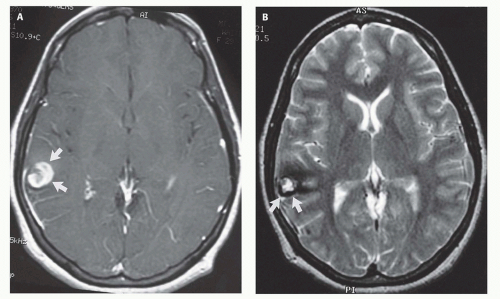
FIG. 12-9. Cavernous malformation. A and B. Child with epilepsy. Contrast-enhanced axial T1-weighted image (A) shows an area of mixed signal intensity surrounded by a low-intensity rim (arrows) in the right posterior frontal lobe. This is the classic appearance for cavernous malformations. Axial T2-weighted image (B) demonstrates T2 hypointensity compatible hemosiderin (arrows) from prior hemorrhage surrounding an irregular hyperintense core.

Some authors have proposed a system for grading cavernous malformations, based upon the signal intensity within the lobules of the mass (118). They postulated that some grades of cavernomas have a higher propensity for hemorrhage than other cavernomas. However, other authors have found no relationship between the grade of cavernoma and the incidence of hemorrhage (119). Kim et al. (120) classified lesions into four types (Table 12-2) based on MRI characteristics recently used to stratify patients into treatment groups. Surgery was performed for the following reasons: histological confirmation when in
question, decompression for mass effect causing neurological deficits, and intractable or chronic seizures (121).


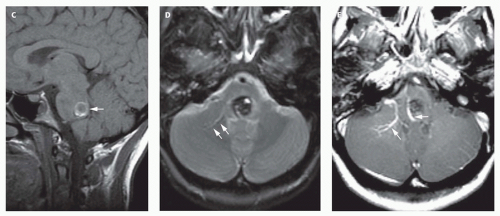
FIG. 12-9. (Continued) C-E. Child with new ocular dysmotility. Axial T1-weighted image (E) shows a hyperintense rim in the dorsal pons (white arrow). Axial T2-weighted image (F) shows the lesion to be heterogeneous, mostly hypointense with areas of intermediate and marked hyperintensity. A large vein (arrows) is located in the left cerebellar hemisphere; note the hyperintensity adjacent to the hypointense vein, due to mismapping of signal from slow flow. Postcontrast T1-weighted image (E) shows two large developmental venous anomalies (arrows), with one coursing directly to the right of the cavernoma.


Developmental Venous Anomalies

DVAs (also called venous malformations) consist of a radially oriented collection of dilated medullary or subcortical veins, which are separated by normal surrounding brain parenchyma, that drain into a single, dilated venous structure (122). No arteriovenous shunting is present. DVAs located in the cerebral hemispheres rarely cause symptoms and are most commonly incidental findings disclosed on diagnostic studies obtained for other reasons. Rarely, DVAs cause clinical symptoms, most commonly headache, focal neurologic deficit, or seizure (123), probably as a result of narrowing of the vein near its site of drainage into a dural venous sinus. This narrowing can result in venous hypertension and, if the draining vein undergoes thrombosis, hemorrhage (124). If the venous hypertension is longstanding, chronic venous ischemia can develop in the region of brain drained by the malformation, sometimes resulting in localized atrophy and resultant seizures. These findings are exceedingly rare in children. DVAs located in the brainstem or cerebellum have a slightly increased incidence of hemorrhage (125), but this is usually the result of rupture of an adjacent cavernoma.


TABLE 12-2 Categories to Characterize Cavernous Malformations Using Spin-echo Sequences on MRI Correlated With Surgical Outcomes


T1 Weighted


T2 Weighted


Margin Characteristics


Type I


Hyperintense


Hyperintense


Mixed


Type II


Mixed


Reticulated


Hypointense


Type III


Hypointense ± hyperintense core


Hypointense


Type IV


Hyperintense extending beyond hypointense rim


Hypointense


From (120).


The MR appearance of venous malformations is quite specific and is identical in children and in adults. A “tuft” of radially arranged, small vessels or “caput medusa” (resembling writhing serpents projecting from the cranium of the mythological demon, Medusa) coalesces to form a single large collecting vessel that drains into a venous sinus (Figs. 12-9 and 12-10). The vessels show uniform enhancement after administration of paramagnetic contrast (126). Because the vessels have relatively slow flow, the signal from the flowing blood may be slightly mismapped in obliquely running vessels, such that a congruent hyperintensity is seen adjacent to the hypointense curvilinear vessel (Fig. 12-9D). The most common locations are the frontal lobes (40%), adjacent to the frontal horn of the lateral ventricle and draining into the longitudinal caudate vein, and the posterior fossa (20%), adjacent to the fourth ventricle and draining into the anterior transpontine
vein, the lateral transpontine vein, or the vein of the lateral recess of the fourth ventricle (127). The parietal lobe is also a fairly common site, accounting for about 15% of venous malformations; the temporal and occipital lobes are less common locations (126,127,128,129). If longstanding venous ischemia has been present, the region of brain that has been drained by the venous malformation may become atrophic and calcified. In approximately 5% of these cases, there may be complicating features such as aneurysms or transitional features including arteriovenous shunting that is the hallmark of a pial AVM. In patients with progressive neurological symptoms or hemorrhage, catheter angiography may be useful to evaluate for high-risk features (130).


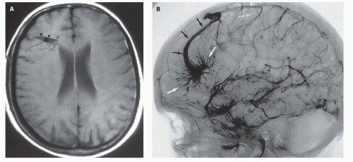
FIG. 12-10. Developmental venous anomaly. A. Axial T1-weighted image shows a tuft of vessels (open arrows) feeding into a curvilinear vascular channel (solid arrows) that drains into the superior sagittal sinus (not seen on this image). This appearance is characteristic of developmental venous anomalies. B. Venous phase angiogram, lateral projection, from a right internal carotid artery injection. The tuft of vessels (white arrows) is seen to form a curvilinear venous channel (black arrows) that drains into the superior sagittal sinus. The findings are identical to what was seen on the MR study.


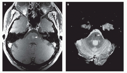
FIG. 12-11. Capillary telangiectasia. A. Axial postcontrast T1-weighted image shows a small area of enhancement (arrowhead) in the middle of the pons. B. Axial T2*-weighted gradient-echo image shows hypointensity of the enhancing region in the central pons (arrowhead), indicating the presence of chronic hemorrhage and establishing the diagnosis of capillary telangiectasia.


Capillary Telangiectasias

Capillary telangiectasias are collections of dilated capillaries separated by normal brain parenchyma likely representing a type of vascular choristoma. They are most commonly found in the pons, rarely bleed, and are usually discovered incidentally at autopsy. Occasionally, they may be found incidentally on routine MRI. Noncontrast scans may be normal, may show slight hypointensity on T1-weighted images, or may show slight hyperintensity on T2-weighted images; no mass effect is present. After administration of paramagnetic contrast, moderate enhancement with slightly brush-like borders is seen (Fig. 12-11). Marked
hypointensity is typically seen on gradient-echo images (Fig. 12-11) (131), presumably secondary to the presence of slowly flowing, deoxygenated blood within the vessels (132).


EXTRADURAL VASCULAR MALFORMATIONS AND NEOPLASMS REQUIRING ENDOVASCULAR TREATMENT

Vascular malformations of the head and neck can be classified in a similar manner to intracranial vascular malformations. There are some distinct differences, however. The histology of the abnormal vessels (arteriovenous, venous, capillary, lymphatic or mixed capillarylymphatic) and the flow characteristics at arteriography (low or high) have been used for categorization (133). Arteriography is used only when the diagnosis is in question or when intervention is contemplated. In addition, certain pediatric neoplasms can induce significant neovascularity resulting in spontaneous hemorrhage or requiring endovascular embolization prior to excision. The presentation and imaging characteristics of common pediatric head and neck tumors are described in Chapter 7. Discussion in this section focuses on the role of endovascular treatment modalities.


Hemangioma

Hemangiomas are benign neoplasms of endothelial proliferation, occurring in up to 10% of children by 1 year of age. Manifestations appear shortly after birth and consist of development of a reddish lobulated mass on the skin. Growth may be rapid during the first year of life. The diagnosis is usually made by clinical examination. Management is usually medical and includes steroids and other immunomodulatory and antiangiogenic agents (134,135,136). Hemangiomas usually stabilize after the first year of life; many will involute and even resolve during the subsequent few years. Atrophy of cutaneous tissues with scar formation may accompany regression of the lesion. Features requiring more aggressive treatment include functional impairment (impingement on the eyelid, lip, or mouth impairing development of vision, feeding patterns, or language, respectively), hemorrhage, mass effect upon the airway, platelet sequestration resulting in coagulopathy (Kassabach-Merrit syndrome) or high output CHF (137,138).

Imaging features of hemangiomas are described in Chapter 7. On angiography, hemangiomas are high-flow capillary lesions with wellcircumscribed areas of dense, prolonged opacification. The feeding arteries are dilated and rapidly drain into dilated veins (139). Intracranial and orbital structures should always be evaluated in patients with hemangiomas of the face or neck, as a subgroup of patients with hemangiomas have the PHACES syndrome (see Chapter 6), a constellation of posterior fossa brain malformations, hemangiomas, arterial anomalies, cardiovascular anomalies, eye abnormalities, and sometimes sternal clefting (136,140,141,142) (Fig. 12-12). When intervention is indicated, surgical resection, laser coagulation, and embolization are potential methods (143,144). Endovascular treatment is generally reserved for lesions causing thrombocytopenia and bleeding diathesis. Such lesions often have a more aggressive histology resembling hemangioendothelioma.

Only gold members can continue reading. Log In or Register to continue

Stay updated, free articles. Join our Telegram channel

Jun 19, 2016 | Posted by in NEUROLOGICAL IMAGING | Comments Off on Anomalies of Cerebral Vasculature: Diagnostic and Endovascular Considerations

Full access? Get Clinical Tree

Get Clinical Tree app for offline access