Congenital Anomalies of the Spine


Congenital Anomalies of the Spine


Erin Simon Schwartz

A. James Barkovich


Normal and Abnormal Embryogenesis of the Spine: An Overview

An understanding of the normal embryonic developmental sequence of the spine is invaluable to the understanding of spinal anomalies. Furthermore, combining knowledge of embryology with understanding of the anatomic relationships in the mature congenital lesion allows insight into the time and stage of development at which the normal sequence was altered; this insight leads to a better understanding of developmental lesions. Normal development of the spine will therefore be discussed in some detail.


Gastrulation and Neurulation

On about the 15th day of embryonic life, ectodermal cells proliferate to form a plate, the primitive streak, along the surface of the embryo. A rapidly proliferating group of cells forms at one end of the primitive streak; this nodular proliferation, surrounding a small primitive pit (known as the Hensen node), defines the cephalic end of the primitive streak. At days 15 and 16, cells enter the primitive pit and migrate inward between the ectoderm and endoderm and then course laterally and undergo epithelial to mesenchymal transition, to form the interposed mesoderm. Initially, no cells migrate in the midline; later, these mesodermal cells will join in the midline to form the notochordal process, which will eventually roll into a tube, separate from the endoderm, and become the notochord. During a process known as intercalation, the notochordal process fuses with the endoderm, creating a communication of the central canal of the notochordal process with the yolk sac. As the central canal is already in communication with the amniotic cavity through the primitive pit, a transient communication is present all the way from the yolk sac to the amniotic sac; this communication is called the primitive neurenteric canal. Once formed, the notochord induces the formation of a plate of ectodermal cells in the dorsal midline, beginning immediately cephalad to the Hensen node (Fig. 9-1). Under the influence of bone morphogenetic proteins (BMPs), most of the ectoderm is prevented from differentiating into neuroectoderm (1). However, suppression of the BMPs by antagonists such as chordin, noggin, and follistatin, emanating from the primitive node, allows neuroectoderm to form in the midline (1,2). The lateral edges of this midline structure, now known as the neural plate, are contiguous with the ectoderm from which the plate has differentiated, now known as superficial or cutaneous ectoderm.

After the neural plate is formed, it is shaped into an elongated structure that is broad at the anterior (cranial) end and narrow at the posterior (caudal) end. The major driving force of the shaping is a mediolateral elongation of a group of cells, along with development of
polarized cellular protrusions that enable the cells to migrate medially and intercalate with neighboring cells close to the midline (3). This midline convergence of cells causes an anteroposterior elongation and narrowing of the neural plate (4,5). This process is strongly related to the development of cell polarity (4,5). Along with the process of shaping, the neural plate also bends. At approximately 17 days of gestation, the lateral portions of the neural plate begin to thicken bilaterally, forming the neural folds; the process of bending elevates these folds and brings them to the dorsal midline (6). The process involves the formation of “hinge points” at two sites: the median hinge point (MHP) in the ventral midline and extending over the rostrocaudal extent of the neural plate and the paired dorsolateral hinge points (DLHPs), which form mainly at the levels of the developing brain (2) and lower spine. The formation of these hinge points is controlled by secretion of the signal transduction protein sonic hedgehog by the notochord as well as an inhibitory interaction between BMP2 and Noggin, particularly in the lower spine (7). After formation of the hinge points, the more lateral aspects of the neural plate are elevated around the MHP, bringing the DLHPs upward and toward the midline (Fig. 9-2). This elevation is accomplished by a poorly understood process called apical constriction, in which columnar cells of the neural tube are converted into wedge-shaped cells (8). Eventually, the lateral folds contact one another in the dorsal midline and adhere to one another, with their fusion forming the neural tube (neurulation). This midline contact (also called neural fold apposition) results from constriction of the open posterior neural tube, which is biomechanically coupled to the zippering point by an F-actin network (9). Neurulation seems to begin separately at least two different levels in humans, when cellular protrusions (possibly cilia) project medially from the most dorsal cells of the neural folds on either side. A third site of closure at the caudal end of the embryo has recently been identified in mouse embryos; if present in humans, this may account for the high incidence of spina bifida at this level (9). Cell recognition and adhesion occur under the influence of many molecules (Ephrin-A5, EphA7, neural cell adhesion molecule, and neural cadherin among them (3)), closing the tube at each point. Immediately following closure, the overlying ectoderm separates from the neural tissue and the edges of the ectoderm meet in the midline and fuse, forming a continuous ectodermal covering of the neural structures, with the mesenchymal cells of the neural crest migrating between the cutaneous and neural ectoderm layers (2). Progressive folding and closure of the neural structures and separation from ectoderm then proceed both cranially and caudally from each point of initial closure, ultimately resulting in complete closure (10,11). The initial closure of the neural tube in humans, the posterior neuropore, is believed to be at the hindbrain-cervical junction, from which closure extends in both directions. A second site of closure in mice is at the forebrain-midbrain boundary; this site has not been confirmed in humans. The necessity of this site even in mice is questionable, as about 80% of mice that lack the second closure point still achieve complete cranial closure (12). The “third” site of closure occurs at the most rostral extremity of the forebrain, the lamina terminalis (2). The exact site of the most caudal end of the neurulation-formed neural tube has been debated, but most experts believe that it is at the S2 level (10,11). Others point out, however, that neural tube defects are not restricted to any specific location(s) and propose that the human neural tube initially closes at a single site with closures extending from that location (13).


Figure 9-1 Schematic diagram showing the development of the neural plate. The primitive streak forms along the surface of the embryo by about 15 days of embryonic life. A small primitive pit lies at what will become the cephalic end of the primitive streak; a nodular proliferation of cells surrounding the primitive pit becomes known as the Hensen node (A, seen from above and B, midline cut of A). On about days 15 and 16, cells enter the primitive pit and migrate cephalad in the midline to form the notochordal process, which will eventually become the notochord. The notochordal process and notochord induce formation of a plate of ectodermal cells dorsally in the midline; this is the neural plate (C, seen from above and D, midline cut of C).

Recent work has shown marked complexity of the process of neural tube closure, involving cellular events such as convergent extension (a fundamental and conserved collective cell movement that forms elongated tissues during embryonic development (14)), apical constriction (constriction of the apical surfaces of the cells in prospective anterior portion of the dorsal visceral ectoderm (8)), and interkinetic nuclear migration (15), as well as precise molecular control via the noncanonical Wnt/planar cell polarity pathway, Shh/BMP signaling, and the transcription factors Grhl2/3, Pax3, Cdx2, and Zic2 (15,16,17). In mammals, this process is regulated by more than 300 genes (18); biomechanical inputs into neural tube morphogenesis have also been identified. Nutritional factors are also important; several rodent studies show that folic acid reduces neural tube closure defects (NTDs) and others have shown that inositol reduces NTDs in some strains of mice in which folate does not work (19). No definitive results have been published at the time this chapter was being written (19). Here,
we review these cellular, molecular, and biomechanical mechanisms involved in neural tube closure, based on studies of various vertebrate species, focusing on the most recent advances in the field.


Figure 9-2 Normal and abnormal neurulation. A-E. Normal neurulation. The neural plate is composed of neural ectoderm, which is continuous with cutaneous ectoderm on either side. The cells at the junction of the neural ectoderm and cutaneous ectoderm will eventually differentiate into neural crest cells (A). At approximately 17 days, the lateral portions of the neural plate begin to thicken, forming the neural folds (B). Contractile filaments located in the neuroepithelial cells in the neural folds contract, causing the neural folds to bend dorsally along the entire length of the neuroaxis, bringing the edges of the neural folds toward one another in the midline (C). Neurulation (closure of the neural tube) begins when the neural folds meet in the midline. At the time of closure, the overlying ectoderm separates from the neural tissue and fuses in the midline, forming a continuous ectodermal covering of the neural structures. At the same time, the neural crest cells are extruded from the neural tube to form a transient structure immediately dorsal to the tube (D). Eventually, these neural crest cells will migrate to form dorsal root ganglia and multiple other structures (E). F-G. Abnormal neurulation. When there is premature disjunction of neural ectoderm from cutaneous ectoderm, the surrounding mesenchyme gains access to the inner surface of the neural tube. When mesenchyme comes in contact with this primitive ependymal lining, it evolves into fat. This is believed to be the process underlying the formation of spinal lipomas (F). Complete nondisjunction of cutaneous ectoderm from neural ectoderm results in the formation of myelomeningoceles (Fig. 9-4). Focal nondisjunction results in a persistent epithelium-lined connection between the central nervous system and the skin (G). This persistent connection has been labeled a dorsal dermal sinus.


Canalization and Retrogressive Differentiation

A portion of the neural tube develops caudal to the posterior neuropore. This elongation is not due to primary neurulation, but is the result of another process known as canalization (or secondary neurulation). In this process, a caudal cell mass (also called the tail bud), composed of undifferentiated, pluripotential cells (the residua of the primitive streak (2,20)), forms in the tail fold as a result of fusion of neural epithelium (at the caudal end of the embryo) with the notochord. Ventral to the caudal cell mass, with the notochord interposed, lies the cloaca, which will form the cells of the anorectal and lower genitourinary tract structures.

By the time the embryo is 30 days old, multiple microcysts and clumps of cells begin to appear in the caudal cell mass. These microcysts coalesce to form an ependyma-lined tubular structure that unites with the neural tube above (Fig. 9-3). This process is not as organized as the process of neurulation; the multiple accessory lumina and ependymal
rests in the normal filum terminale and distal conus medullaris of the adult are believed to result from the disorder of this process. Several neuronal markers expressed in vertebrate embryos are thought to modulate the differentiation of structures derived from the caudal cell mass and be involved in the maturation of the caudal spinal cord. These include N-CAM, synaptophysin, 3A10, and NeuN (21). The final stage in the formation of the distal spinal cord begins at about 38 days of gestation, at which time the cell mass and central lumen of the caudal neural tube decrease in size as a result of programmed cell death (apoptosis) of the portion derived from primary neurulation (20) and necrosis of the portion derived from secondary neurulation (22). This process has been named retrogressive differentiation (Fig. 9-3). The caudal segment (formed by canalization and retrogressive differentiation) eventually becomes the most caudal portion of the conus medullaris, the filum terminale, and a focal dilation of the central canal (within the conus medullaris) known as the terminal ventricle (ventriculus terminalis) (23,24,25,26,27,28).


Figure 9-3 Canalization and retrogressive differentiation. After formation of the neural tube, a caudal cell mass forms in the tail fold as a result of fusion of neural epithelium at the caudal end of the embryo with the notochord (A). By the age of 30 days, multiple cysts and clumps of cells appear in the caudal cell mass (B). These cysts coalesce to form a tubular structure that unites with the neural tube above (C). At about 38 days, the cell mass and central lumen of the caudal neural tube decrease in size through cell necrosis in a process known as retrogressive differentiation. The segment formed by this process eventually forms the distal-most conus medullaris, the filum terminale, and the terminal ventricle (D).


Formation of the Vertebral Column

Formation of the vertebral column was discussed in Chapter 2 and illustrated in Figure 2-21. A review of the subject is given here, as well, for convenience of the reader. Development of the vertebral column can be divided into three periods. The first of these is membranous development. At about the 25 gestational day, the notochord separates from the primitive gut and neural tube to create two zones, the ventral subchordal and dorsal epichordal zones. These zones are filled with mesenchyme, which migrates to them from its initial position lateral to the neural tube.

The mesenchyme lateral to the closing neural tube organizes into somites, which are separated by small intersegmental fissures. Somite formation progresses from rostral to caudal (29). Each somite is divided into a medial (medial sclerotome) and a lateral (lateral myotome) portion. The medial sclerotome contributes to the formation of the vertebrae, while the lateral myotome gives rise to the paraspinous musculature. After the neural tube has closed and becomes separated from the superficial ectoderm, mesenchyme also migrates dorsal to the neural tube to form the precursors of the neural arches (in addition to the meninges and paraspinous muscles). Differentiation of the ventral and dorsolateral mesenchyme, which will form the vertebral body, results from induction by the sonic hedgehog protein, whereas the differentiation of the dorsal mesenchyme, which will form the posterior elements, results from induction by a protein called BMP4 (30). Subsequently, the sclerotomes separate transversely along the previously mentioned intersegmental fissures. The inferior half of one sclerotome then fuses with the superior half of the subjacent sclerotome across the fissure to form a vertebral body. This process proceeds bilaterally and symmetrically so that the fusion of the sclerotomes on each side forms the half of the vertebral body on that side. As a consequence of this resegmentation, the intersegmental arteries and veins become located in the center of the new vertebral bodies.

During the second (chondrification) stage of vertebral development, chondrification centers appear within sclerotomes at the cervicothoracic junction region during the sixth embryonic week and then extend rostrally and caudally. Six centers will form each vertebral level, with two in the vertebral centrum, two forming the posterior vertebral arches and spinous process, and two within the transverse processes and costal
arch (31). Notochordal remnants persist between the newly formed vertebral bodies and become incorporated into the intervertebral discs as the nuclei pulposi. Portions of the thoracic sclerotomes later migrate ventrolaterally to form ribs. The anterior and posterior longitudinal ligaments form during chondrification, from mesodermal cells.

In the final stage of vertebral development (ossification), the chondral skeleton ossifies to complete the formation of the vertebrae. Ossification starts in three centers, one in the middle of the vertebral body and one in each vertebral arch. The thoracolumbar junction region is the first to ossify, with rostral and caudal ossification to follow. The formation of the vertebrae at the caudal end of the embryo proceeds by a different, less organized process. A mass of cells composed of notochord, mesenchyme, and neural tissue merely divides into somites to form the sacral and coccygeal levels. Regression results in reduction and fusion of most of these segments. As in development of the caudal neural tube, this apparent disorganization of the caudal cell mass leads to frequent anomalous development. Caudal regression syndromes, lipomas, and teratomas can result (32,33).


Anomalies of Spinal Formation: Concepts


Embryological Concepts and Theories

Embryological explanations of anomalies of the developing spine are continually evolving. As new facts are discovered, old theories sometimes have to be discarded and new theories developed. In contrast to mathematical theories, embryological theories cannot be proved correct; they can only be verified or disproved. In clinical medicine, a theory is useful if it explains observations and helps to organize a classification; the ability to predict future observations is an added bonus. The classification scheme used in this chapter is based primarily upon a concepts developed in the early 20th century by Della Rovere (34), modified by David McLone, MD, PhD., and Thomas Naidich, MD in the 1980s, by Tortori-Donati and colleagues (35) in the 1990s and most recently by Andrew Copp and colleagues at University College in London (15). These more recent discussions include new genetic, transcriptomic, biochemical, molecular pathway and environmental information that answers some questions but raises others. This chapter will discuss various aspects of all classifications but will focus on the more recent concepts.

Developmental neurogenetics are largely ignored in the discussion of spine anomalies in this chapter. This is not in any way an attempt to minimize the value of developmental neurogenetics. Indeed, spinal anomalies result from a multitude of factors and genetic factors are likely among the most important (interested readers should start with the most recent reviews) (15,17,18). However, despite many major advances in genetics over the past 5 years, the details of developmental neurogenetics of normal and abnormal spinal development are still being investigated and the inclusion of the bits and pieces that are known would likely prove more confusing than enlightening to most practicing clinicians.


Neurulation Anomalies

The process of separation of the neural tube from the cutaneous ectoderm during closure of the neural tube is known as disjunction. After disjunction, the cutaneous ectoderm fuses in the midline, dorsal to the closed neural tube. At the same time, perineural mesenchyme migrates into the newly created space between the neural tube and cutaneous ectoderm, surrounds the neural tube, and is induced to form the meninges, the bony spinal column, and the paraspinous musculature (36). The mesenchyme normally remains isolated from the newly formed central canal of the spinal cord because the neural tube closes immediately prior to, or simultaneously with, disjunction. Recently, a number of papers have found suggestions that gene dysregulation associated with both neuronal development and axonal growth and guidance (37) and loss of protein function (38), inflammatory factors (37), and epigenetic factors such as histone 3 levels and protein methylation (39,40) are important factors in the growth and closure of the spinal cord. As these functions are investigated and better understood, it is likely that our knowledge of these factors will change our approach to the treatment of neural tube defects.

Whatever the genetics and posttranscriptional modifications involved in neural tube development, defective disjunction can explain a number of diverse pathologic lesions. For example, focal unilateral premature disjunction of the neural ectoderm from the cutaneous ectoderm (prior to closure of the neural tube) allows the perineural mesenchyme to gain access to the neural groove and come in contact with the primitive ependymal lining of the groove. Mesenchyme that is exposed to the interior of the neural tube seems to develop into fat, either by receiving signals promoting adipocyte (fat cell) production or by the blockage of signals that prevent adipocyte production (41,42). Therefore, focal premature disjunction of neural ectoderm from the superficial ectoderm could explain the development of spinal lipomas and lipomyelomeningoceles (43) (Fig. 9-2F). Spinal lipomas situated rostral to the filum terminale seem to be formed in this manner. Lipomas caudal to the conus medullaris more likely form as a result of other processes, such as abnormal development of the caudal cell mass.

Other anomalies of the spine can be explained by failure of disjunction. Dermal sinus tracts may be the consequence of focal failure of disjunction, resulting in a focal ectodermal-neural ectodermal tract (Fig. 9-2G). A related entity has been recently described, the dermal sinus-like stalk, in which there is a solid stalk composed of fibrous tissue and fat (with or without nervous tissue) rather than the epithelial-lined tract of the typical dermal sinus (44). The tract should prohibit mesenchyme from migrating between the neural ectoderm and cutaneous ectoderm at that site, forming a focal spina bifida as is commonly seen in children with dermal sinus tracts. Moreover, the adhesion between the ectoderm and neural ectoderm could explain the correlation of the dermatome level of the cutaneous lesion with the neuroectodermal level of the central nervous system termination of the tract. Open spinal dysraphism (OSD) (myelocele and myelomeningocele) can be explained a large area of nondisjunction (Figs. 9-2 and 9-4). These concepts will be discussed in more detail in later sections of this chapter.


Association of Spinal Anomalies with Other Systemic Anomalies

Spinal malformations are very commonly associated with anomalies of the viscera, as well as those of the spinal column. As discussed previously, the caudal cell mass forms in close anatomic proximity to the cloaca, the region of origin of the lower genitourinary tract and anorectal structures. As a result of this close embryological relationship, patients with anorectal and urogenital anomalies have a high incidence of lumbosacral hypogenesis, terminal myelocystoceles, and tethered spinal cords (and vice versa) (45,46,47). Moreover, the notochord is associated with the induction of normal formation of thoracic and abdominal viscera, as well as the neural tube. Therefore, patients with spinal anomalies secondary to abnormal formation of the notochord will often have anomalies of the upper gastrointestinal tract or the respiratory tract. Finally, it is important to remember that the development of the vertebral column is influenced by many of the same factors that influence development of the spinal cord. Therefore, any condition that results in vertebral anomalies, such as the VATER syndrome (48), the Klippel-Feil syndrome (49), and lumbosacral hypogenesis (50,51) should be investigated for associated anomalies of the spinal cord.


Figure 9-4 Open spinal dysraphism (myelocele and myelomeningocele). A. Myelocele. The neural placode is a flat plaque of neural tissue that is exposed to the air. The dura is deficient posteriorly; the pia and arachnoid line the ventral surface of the placode and dura, forming an arachnoid sac that is continuous with the subarachnoid space superiorly and inferiorly. Both the dorsal and ventral roots arise from the ventral surface of the placode. B. Myelomeningocele. This is identical to the myelocele with the exception that there is an expansion of the ventral subarachnoid space, which posteriorly displaces the placode.


Clinical Manifestations of Spinal Anomalies

Many patients with spinal anomalies present either with external manifestations of the anomaly or with the tight filum terminale (tethered cord) syndrome. The external manifestations vary with the type of anomaly and are described in more detail in the sections on the specific anomalies themselves. They include cutaneous hemangiomas, dimples, hairy patches, atypical scoliosis, and large subcutaneous lipomas. The syndrome of the tight filum terminale, also known as the tethered spinal cord syndrome, denotes a complex of neurologic and orthopedic deformities. This clinical syndrome may be associated with split-cord malformation (SCM), spinal lipoma, and syringomyelia or with a short, thick filum terminale; frequently, the conus medullaris is in a low position. Patients may present with new onset of symptoms at any age (52); the authors have seen patients in nearly every decade of life (as late as the eighth decade) become suddenly symptomatic from their tethered spinal cords. Patients suffer from difficulty with locomotion, ranging from muscle stiffness to actual weakness; some exhibit abnormal lower extremity reflexes (53). The patients can also exhibit bladder dysfunction (usually manifest as a low-pressure, dribbling urine stream), sensory changes with abnormal somatosensory-evoked potentials, orthopedic deformities of the lower extremities (most commonly clubfoot), and back pain (particularly with exertion). Urodynamic studies are typically abnormal. Bowel dysfunction is uncommon. Although scoliosis frequently accompanies this syndrome, it is rarely the sole complaint (53,54). Symptoms are frequently worse in the mornings and after exercise. An increased incidence of tethered spinal cords is seen in lumbosacral hypogenesis, in the VATER syndrome (48,51), and in anorectal malformations (imperforate anus), including low lesions (55). If the condition is recognized and treated early, urologic and motor outcomes are generally good (53,56).

Adults with tethered spinal cords may present somewhat differently than affected children. Adults present more often with pain, possibly because of accompanying degenerative changes, and less commonly with incontinence, weakness, or scoliosis (52,57). Indeed, it has been suggested that tethering of the spinal cord accelerates disc degeneration. Adults who present with symptoms of lumbar disc degeneration without MRI evidence of disc disease should be investigated for a tight filum terminale (58).


Terminology

The term spinal dysraphism refers to a heterogeneous group of spinal anomalies. In spite of their heterogeneity, all lesions within this group have incomplete midline closure of mesenchymal, osseous, and nervous tissue (59).

Spina bifida refers to incomplete closure of the bony elements of the spine (lamina and spinous processes) posteriorly (60).

Open spinal dysraphism (OSD) includes the myelocele, a midline plaque of neural tissue that lies exposed at and is flush with the skin surface, and the myelomeningocele, a myelocele that has been elevated above the skin surface by expansion of the subarachnoid space ventral to the neural plaque.

Closed spinal dysraphisms are a group of lesions that develop beneath an intact dermis and epidermis; that is, there is no exposed neural tissue. The clinical-radiological classification system of Tortori-Donati, Rossi, and Cama subdivides this group into lesions with a subcutaneous mass (usually the result of a subcutaneous lipoma or a simple meningocele) and those without a subcutaneous mass (35) (Table 9-1). Included in the category of closed spinal dysraphism with a subcutaneous mass are lipomas with dorsal defect of the bony spinal canal (lipomyelocele and lipomyelomeningocele), meningocele, and myelocystocele. Anomalies with closed spinal dysraphism lacking a subcutaneous mass include
the simple dysraphic states (fatty and/or fibrous thickening of the filum terminale, intraspinal lipoma, spina bifida, and persistent terminal ventricle) and the complex dysraphic states (split-cord malformation, dorsal dermal sinus, caudal regression syndrome, segmental spinal dysgenesis, neurenteric cyst, and dorsal enteric fistula).


Table 9-1 Clinical-Radiological Classification System for Spinal Dysraphism


Open Spinal Dysraphism


(Hemi)myelomeningocele


(Hemi)myelocele (also known as myeloschisis)


Closed Spinal Dysraphism


With a subcutaneous mass


Lipoma with dorsal defect (lipomyelomeningocele and lipomyelocele)


Myelocystocele (terminal or cervical)


Meningocele


Cervical myelomeningocele


Without a subcutaneous mass


Simple Dysraphic States


Spina bifida


Persistence of the terminal ventricle


Intraspinal lipoma (intradural or filum terminale)


Tight filum terminale (fibrous thickening)


Complex Dysraphic States


Dorsal dermal sinus


Caudal regression syndrome


Split notochord syndrome (dorsal enteric fistula and neurenteric cyst)


Split-cord malformation (diastematomyelia and diplomyelia)


Segmental spinal dysgenesis


Adapted from Tortori-Donati P, Rossi A, Cama A. Spinal dysraphism: a review of neuroradiological features and embryological correlations and proposal for a new classification. Neuroradiology 2000;42:471-491.


Counting Anomalous Vertebrae

Identifying the specific level in patients with vertebral anomalies, such as hemivertebrae and butterfly vertebrae, can be challenging. One way to approach this is to count all of the vertebrae (normal ones, butterfly vertebrae, and hemivertebrae) in the numbering, but to designate the hemivertebra as being extra by adding an “h” and the butterfly by adding a “b.” For example, if C6 is a hemivertebra, it is referred to as C6h. A butterfly T8 would be T8b. A report of a patient with these two segmentation anomalies would state that the spine is not a C7/T12/L5/S5 (i.e., normal) spine but a C8(1h)/T12(1b)/L5/S5 spine with the hemivertebra at C6 and the butterfly vertebra at T8. This system can be useful for transmitting the necessary clinical data to the referring physician. Accurate determination of vertebrae can only be performed by labeling from the craniocervical junction and proceeding inferiorly. Recently, reported lumbar level determination utilizing L5 nerve root morphology has been shown in adults to be over 98% accurate; however, this technique remains to be validated in children (61).


Imaging Techniques

The spine can be imaged using a variety of techniques. Sonography and magnetic resonance imaging (MRI) can all give exquisite images of the diverse anatomic anomalies that characterize these entities (62,63,64,65,66,67); sonography is most useful, as expected, in neonates and young infants when the vertebrae are smaller and nonossified. CT may be needed occasionally for detailed information about osseous structures, but it is very uncommon. At UCSF and CHOP, MRI is imaging modality most commonly used for the diagnosis and presurgical planning of these entities. For fetal imaging, MRI and ultrasound are complimentary, and both are usually necessary for a thorough evaluation (68).

An important concept to remember is that a single patient may have multiple spinal anomalies. For example, a patient with a lipomyelomeningocele may have, in addition, a split-cord malformation, a tight filum terminale, or a dorsal dermal sinus. Therefore, it is important to image the entire spine if a cutaneous anomaly or vertebral anomaly suggests the presence of a malformation. The ability to noninvasively perform high-resolution imaging of the entire spine in the sagittal and coronal planes in a very few sequences without ionizing radiation is an advantage of MR that other modalities cannot match. For example, one study comparing MR and ultrasound in the infant spine found that, while a normal ultrasound was sufficient to rule out most spinal pathology, additional pathology is picked up by MR in 20% of cases (69). Detection of a thick or fatty filum terminale in a patient whose conus medullaris terminates at a normal level may be difficult with sonography but is easy on axial MR images. Finally, sonography becomes progressively less useful as ossification of the posterior elements proceeds during the first year of life while MR remains superb. Typical MR sequences for spine imaging are discussed in Chapter 1. It should be added that spiral CT, with the rapid scanning time and easy curvilinear and planar reformations (in any plane), allow excellent evaluation of osseous spinal anomalies and, if intrathecal contrast is administered, good evaluation of intraspinal lesions. In our practices, MR is currently the initial imaging modality of choice because it is noninvasive and does not use ionizing radiation, but for some cases, CT myelography gives important supplementary information. The addition of submillimeter steady-state (FIESTA, CISS) or T2 RARE volumetric imaging through the lumbosacral thecal sac may permit better characterization of anomalies, including improved visualization of nerve roots (70,71).


Imaging of Patients with Idiopathic Scoliosis

While the necessity of preoperative imaging with MRI was previously controversial, there is now general agreement that patients with scoliosis should have their spinal columns imaged by MR before undergoing distraction and instrumentation of their spines (72). This is particularly true in any patient presenting with clinical deterioration, atypical features of scoliosis (rapid progression, abnormal location of the curve, or a left thoracic curve), neurological signs or symptoms (73), age less than 11 years (74), or severe scoliosis (Cobb angle > 45°-50°) (73,75). In these patients, MR should be used to look for neoplasm, Chiari I deformity, syringohydromyelia, tethered spinal cord or other cord anomaly, and intra- or extradural cysts. Any of these conditions can lead to scoliosis; more importantly, surgical treatment of the scoliosis before treatment of the underlying disorder may result in increased neurologic disability.


Abnormalities of Neurulation (Disorders in Which the Spinal Cord Does Not Completely Fuse Posteriorly)


Disorders Resulting from Abnormal Disjunction


Nondisjunction (Open Spinal Dysraphism: Myelocele and Myelomeningocele)

Myeloceles and myelomeningoceles most likely result from a localized lack of closure of the neural tube, resulting from a lack of expression of specific receptors on the surface of the neuroectodermal cells (15). Most authors use the term “myelomeningocele” to refer to any OSD; we use the term OSD to encompass the myelocele and myelomeningocele. All other anomalies are considered closed spinal dysraphisms.

Open neural tube defects cause neurological disabilities, including paraplegia, hydrocephalus, incontinence, sexual dysfunction, skeletal deformities, and, often, cognitive impairment (76). Experimental evidence suggests that the neurological deficits of patients with OSD are not entirely caused by the open neural tube defect but also by changes in transcriptomics, epigenetic factors, chronic mechanical injury, and amniotic fluid-induced chemical trauma that progressively damages the exposed fetal neural tissue during gestation (15,18,37). The prenatal diagnosis of OSD by ultrasound or MR (Fig. 9-5) has made postnatal diagnosis much less common, in regions where these modalities are widely available. Over the past 15 years, in utero OSD repairs in human fetuses have shown significant improvements in outcomes of children so treated; reports suggest postoperative improvement of the associated Chiari II malformation, ventriculomegaly, lower extremity function, and brain stem function (77,78,79,80) in fetuses treated before 26 gestational weeks. As a result, in utero OSD repair in human fetuses is now the standard of care in the United States, having been shown to significantly reduce the need for ventricular shunting, improve hindbrain herniation, and result in superior motor outcomes in a multicenter, prospective trial (81) in fetuses treated before 26 gestational weeks. Intermediate follow-up of patients having undergone in utero repair has shown sustained improvement in hindbrain herniation, ambulation, and continence (82). Thus, prenatal diagnosis of OSD by ultrasound or MR (Fig. 9-5) has made postnatal diagnosis much less

common, in regions where these modalities are widely available. The ongoing prospective multi-institutional follow-up study will determine the long-term benefits of prenatal repair of this spinal anomaly on neurological and functional outcome.


Figure 9-5 Prenatal imaging of an open spinal dysraphism (myelomeningocele). A. Sagittal sonogram using a 4-MHz transducer shows a focal loss of the normal echogenicity of the posterior elements of the vertebrae, indicating a bony spina bifida. The meningocele is seen as a hypoechogenic region (arrows) extending dorsally at the level of the spina bifida. B. Higher-resolution image using a 8-MHz transducer shows curvilinear echoes (arrows) representing neural tissue running through the meningocele. C. Sagittal image through the spine shows the lumbosacral spina bifida with the dorsal myelomeningocele (small white arrows). The Chiari II malformation (larger white arrows) is seen at the craniocervical junction. Note the massive hydrocephalus. D. Axial image shows the dorsal bony spina bifida. At this level, the placode (black arrows) is still within the spinal canal. E and F. Extensive cerebellar herniation due to myelomeningocele in a different patient. Image (E) shows an open spinal defect/spinal bifida at the S1 level; no meninges were apparent. Image (F) shows a very small posterior fossa; the black arrow shows a nearly vertical tentorium cerebelli, and the white arrow shows cerebellar tissue extending down to the C5 level. G. Sagittal midline MRI of the same patient as in (E and F) at age 4 months, showing the effects of a prolonged severe CSF pressure gradient. The cerebellum is small and irregular in shape; normal folial patterns cannot be seen. The brain stem is elongated and very narrow, with the pontomedullary junction at the level of C1. Cerebellar tissue extends to the bottom of C4 (white arrow).


Figure 9-5 (Continued)

The exact cause of the impaired closure of the neural tube in patients with OSD has not been discovered. Environmental factors implicated in increasing the risk include socioeconomic class, maternal age, maternal diet, maternal diabetes and obesity, and exposure to antiepileptic drugs. Genetic components include association with trisomy 13, 18, and 21; association with genetic syndromes (Meckel syndrome and anal stenosis); racial differences in incidence rates; increased risk for a second affected child (3-5×) for couples who have one affected infant; and a 10-fold increase in risk for siblings of affected children compared to the general population (83,84). Strong evidence has accumulated for a protective role of folate in this disorder. Animal work suggests that the folate deficiency impairs biosynthesis of pyrimidine, the presence of which may help to compensate for an underlying genetic predisposition (85). Localized differences in folate metabolism exist early during neural tube formation, and it is believed that folate may modulate proliferation in the midline via effects on Aldh1l1+ cells (86). Offspring of women whose diet is supplemented with folate have a significantly reduced incidence of neural tube defects (87,88,89,90). This has led to investigations of genes involved in folate metabolism as being potentially related to development of OSD. Two identified polymorphisms (C677T and possibly A1298C) in the homocysteine remethylation gene MTHFR are associated with a 1.8-fold increase in the risk of an open neural tube defect (12). Offspring of obese women (defined as weighing more than 70 kg) have an increased incidence of neural tube defects, possibly because women with high body mass indices have low folate levels compared with nonobese women, even when controlling for folate intake (91).

In patients with OSD, the neural folds do not fuse in the midline to form a neural tube in the affected zone; instead, the neural tube remains open and the neural folds remain in continuity with cutaneous ectoderm at the skin surface (nondisjunction). The region of open spinal cord, located at the skin surface and open to the air in the posterior midline, is a region of reddish tissue referred to as the neural placode. The posterior (dorsal) surface of the placode is made up of the tissue that would normally form the internal, ependymal lining of the neural tube. The anterior (ventral) surface of the placode corresponds to what would normally be the external surface of the spinal cord (pia mater) (Fig. 9-4).

The lack of separation of the placode from the cutaneous ectoderm prevents the mesenchyme from migrating into the area posterior to the neural ectoderm; it is forced to remain anterolateral to the nervous tissue. Thus, the pedicles and lamina (which are formed from this mesenchyme) are everted, facing posterolaterally instead of posteromedially (Fig. 9-4). As a result of the external rotation of the lamina and pedicles, the spinal canal undergoes a fusiform enlargement throughout the extent of the posterior osseous defect. The maximum enlargement of the canal occurs when the laminae are in the sagittal plane; further rotation of the lamina diminishes the size of the canal (59).

The vertebral bodies can be nearly normal or can have anomalies of segmentation ranging from a single hemivertebrae to a jumbled mass of malsegmented vertebral components. Segmentation anomalies result in a short radius kyphoscoliosis in approximately one-third of patients with myelomeningoceles (59). Another 65% of affected patients develop a (less severe) kyphoscoliosis as a result of a neuromuscular imbalance (92).

In children with lumbar or lumbosacral OSD, the spinal cord is always tethered. The nerve roots in affected patients radiate in a spokewheel pattern as they leave the placode and travel rostrally, laterally, and caudally to their respective neural foramina.

Imaging studies for OSD are most commonly performed in the fetus; the defect is often detected in mid or late second trimester. The OSD itself is fairly easily seen by the absence of the posterior elements
of the spinal column at the affected level, by the posterior fossa contents extending caudally through the foramen magnum and, in many cases, by ventriculomegaly. In general, when the OSD is uncovered and opens to the amniotic fluid, hindbrain herniation is more severe because the rostrocaudal pressure gradient is increased (Fig. 9-5E-G). Early (prenatal) repair is very important in these cases to minimize these consequences.

Imaging studies are rarely required in the newborn with an OSD (Fig. 9-6). The exposed placode is obvious upon visual examination and is usually repaired within 48 hours. After repair, the infants typically have a neurological deficit that is stable. They should not further deteriorate if the accompanying hydrocephalus, which is almost always present unless the patient has had a prenatal repair, is controlled. Neuroradiologic examination of the spine is indicated if patients deteriorate neurologically in spite of adequate treatment of hydrocephalus, if they have an unusual neurological examination, such as an asymmetric lower extremity neurological deficit, which should raise suspicion that the patient has a split-cord malformation and hemiOSD.


Figure 9-6 Myelomeningocele in a neonate. A and B. Sagittal T1-weighted (A) and T2-weighted images (B) show the spinal cord coursing caudally within the spinal canal to the sacral level. The cord is expanded by syringomyelia (s), with some air (small white arrows) producing some susceptibility artifact within the syrinx. The neural placode (p) extends dorsally through the bony spina bifida. The asterisks (asterisk) mark a wet cloth covering the placode. C. Axial T1-weighted image shows the spinal cord (arrowheads) traversing the subarachnoid space from the spinal canal to the skin surface where it will form a placode. D. Axial T1-weighted image at a lower level shows the placode (p) at its dorsal extent, covered by the wet cloth (asterisk).


HemiOSD

Cameron (93), Emery and Lendon (94), and Pang (95) showed that between 31% and 46% of patients with OSD have associated split-cord
malformation. The spinal cord may be split above (31%), below, (25%) or at the same level (22%) as the OSD (94). In addition to the patients with frank split-cord malformation, 5% of patients with OSD have a duplication of the central canals of the spinal cord cephalic to and at the level of the placode, indicating a mild form of splitting that is insufficient to affect the gross contour of the cord (94).

The hemiOSD, a special form of OSD with split-cord malformation, is observed in less than 10% of patients with OSD (94,96). In the hemiOSD, one of the two hemicords exhibits a small OSD, which tends to lie on one side of the midline, whereas the other hemicord is either normal, tethered by a thickened filum terminale (Fig. 9-7), or has a smaller OSD at a much lower level. The two hemicords usually lie in separate dural tubes that are separated by a fibrous or bony spur. Occasionally, the two hemicords lie within a single dural tube, which becomes deficient at the level of the hemiOSD. In general, affected patients have impaired neurological function on the side of the hemi-OSD but normal or nearly normal function on the normal side (96). Imaging studies (Fig. 9-7) will show the splitting of the spinal cord, the extent and symmetry (or lack thereof) of the OSD, the presence or absence of the bony spur, and any other anomalies that may alter the surgical approach. Split-cord malformation (diastematomyelia) is discussed more fully later in this chapter.


The Chiari II malformation

The Chiari II malformation is an anomaly of the cervical spinal cord, brain stem, and hindbrain that is observed in varying degrees in all patients with myelomeningoceles. Because of the close association of Chiari II malformations with OSD, a brief discussion is appropriate in this section, in spite of the fact that this malformation is discussed extensively in Chapter 5.

The Chiari II malformation can be best considered as an entity in which the posterior fossa is too small (114) due to collapse of the ventricular system from leakage of CSF through the open neural tube. As a result of the small bony posterior fossa and the rostral-to-caudal pressure gradient resulting from the CSF leakage, normal contents of the posterior fossa are pulled caudally and, as a consequence, distorted as they are squeezed out through an enlarged foramen magnum. The
brain stem is pulled/stretched inferiorly and narrowed in the anteroposterior diameter, often lying at the level of the foramen magnum or in the cervical spinal canal. The cervical spinal cord is displaced inferiorly and the upper cervical nerve roots have to ascend toward their respective neural foramina. The medulla is also displaced inferiorly. In 70% of patients, it folds caudally at the cervicomedullary junction, dorsal to the cervical spinal cord (which is tethered by the dentate ligaments and therefore limited in its vertical descent), forming a characteristic cervicomedullary kink (Figs. 5-178 and 5-179). The cerebellar vermis often herniates inferiorly, forming a tongue of tissue posterior to the medulla that usually extends down to the C2 or C4 level. Rarely, it extends down into the upper thoracic segments of the canal. The cerebellum wraps around the brain stem (Fig. 5-179). The fourth ventricle is vertical in orientation (Figs. 5-178 and 5-179), extending inferiorly between the medulla and cerebellar vermis; occasionally, it extends down below the medulla, posterior to the cervical spinal cord, in a cyst-like fashion. The quadrigeminal plate is stretched posteriorly and inferiorly (Figs. 5-178 and 5-179). The small posterior
fossa and the downward herniation of its contents result in a low-lying, abnormally vertical tentorium cerebelli (115,116,117,118). Many of these changes improve or completely resolve after fetal repair of the OSD, as discussed in Chapter 5.


Figure 9-10 Patient who had myelomeningocele repair at birth, now with worsening neurologic exam secondary to arachnoid scarring/loculations. A. Sagittal T2-weighted image shows the conus medullaris (white arrow) extending caudally to the L5 level. Based on this image, it cannot be determined whether the neurologic symptoms are the result of retethering or another process. This patient emphasizes the need to image the entire spinal cord. The levels of spina bifida (white arrowheads) are those without spinous processes. B. Sagittal T2-weighted image of the cervical and upper thoracic spine shows a thin spinal cord (black arrows) that is dorsally displaced due to scarring and arachnoid loculations. C. Axial T1-weighted image at the midthoracic level shows the spinal cord (white arrows) to have a crescentic shape, being displaced by the scarring and loculations.


Late/Incomplete Disjunction: Dorsal Dermal Sinuses and Sinus-Like Tracts (Also Considered as a Complex Spinal Dysraphism)


Clinical features

Dorsal dermal sinuses are epithelium-lined tubes that extend inward from the skin surface for varying distances and frequently connect the body surface with the central nervous system or its coverings. This anomaly seems to result from a focal area of late and incomplete disjunction of cutaneous ectoderm from neural ectoderm during the process of neurulation (Fig. 9-2). When the spinal cord later becomes surrounded by mesenchyme and undergoes its relative ascent with respect to the spinal column, this adherence remains and forms a long, epithelium-lined tract. The incidence of dermal sinuses is low in the cervical region, where the neural folds first fuse into the primitive neural tube, and relatively great in the lumbosacral and occipital regions, which are the last portions of the neural tube to close. In a series of 120 cases of dorsal dermal sinuses, 1 was sacrococcygeal, 72 were lumbosacral, 12 thoracic, 2 cervical, and 30 occipital; the remaining 10 were mostly ventral to the skull and spinal column (119). Dermal sinuses in the frontonasal region are discussed in Chapter 5.

Dorsal dermal sinuses occur equally frequently in males and females and are usually detected any time from early childhood through the third decade of life. Physical examination reveals a midline (rarely paramedian) dimple or pinpoint ostium that is frequently associated with a hyperpigmented patch, hairy nevus, or capillary angioma (120). The patients become symptomatic either by infection or because of compression of neural structures by an associated inclusion cyst. Meningitis or abscesses in the subcutaneous, epidural, subdural, subarachnoid, and/or subpial spaces may develop as a result of bacterial ascent through the sinus tract (121,122,123); meningitis is a risk factor for poor outcome (124). Occasionally, the meningitis is chemical in nature, resulting from the release of cholesterol crystals or other contents of inclusion cysts into the cerebrospinal fluid (124,125).

The related spinal dermal sinus-like stalk has been reported as a discrete, albeit rare, entity (44,126). These patients do not have a tract with an epithelial-lined lumen communicating with a cutaneous orifice, but rather a solid stalk of connective tissue, fat, and occasionally neural tissue, that runs from a skin dimple without an ostium (usually covered by cutis aplasia, a translucent layer of thin, parchment-like skin (126)) to the intradural space or spinal cord. The dural sleeve associated with the stalk was directed toward the skin surface, unlike the dural sleeve of the dermal sinus, which is directed toward the spinal cord; this observation suggests an etiology distinct from that of the dermal sinus. The authors postulate that disjunction between cutaneous and neural ectoderm occurred, but that intervening mesodermal cells may have formed a tight, persistent connection between the two. Patients with the sinus-like stalks have a different clinical presentation, usually one of a tethered cord (impaired reflexes, sensory deficit, sphincter signs); both infection and neurologic disability due to an associated (epi)dermoid are rare (126,127).


Pathology

Pathologically, dermal sinuses are thin, epithelium-lined channels that course inward from the skin surface through the subcutaneous tissues, extending into the spinal canal in 50% to 70% of cases. The sinus may reach the dura without passing through it; in such cases, the dura and arachnoid are tented dorsally at the attachment of the sinus tract to the dura. This tenting may be the only manifestation of the sinus tract on myelography. If they pass through the dura, the sinuses may empty into the subarachnoid space or may traverse the subarachnoid space to terminate within the conus medullaris, filum terminale, a nerve root, a fibrous nodule on the dorsal aspect of the cord, or an inclusion cyst (Fig. 9-11) (128,129,130). About half of dorsal dermal sinuses end in inclusion cysts. Conversely, 20% to 30% of inclusion cysts are associated with dermal sinus tracts (129,130,131,132,133). Dermal sinuses can be midline or paramedian in location. Those with midline orifices are usually associated with midline dermoid tumors. Paramedian orifices are more commonly associated with epidermoids, which can be located laterally in the epidural, subdural, or subarachnoid spaces. In some patients, the dermal sinus courses horizontally beneath the skin to the dura mater. In others, it courses subcutaneously for some distance before reaching the dura and then ascends within the spinal canal to the level of the conus medullaris. The exact course of the sinus varies from patient to patient. Therefore, the complete course of each dermal sinus must be determined by narrow, contiguous, or volumetrically acquired sections above and below the level of the external orifice.

The bony abnormalities associated with dermal sinuses are variable. Bone abnormalities may be absent if the dermal sinus extends intraspinally through a ligamentous defect at the interspace between two spinous processes. In other cases, the sinus may be associated with a groove in the upper surface of the spinous process and lamina of the vertebra, a hypoplastic spinous process, a single bifid spinous process, focal multilevel spina bifida, or a laminar defect (129,131).


Figure 9-11 Schematic of dorsal dermal sinus. A tuft of hair, nevus, or hemangioma frequently marks the ostium of the sinus. The dura is often tented when the sinus penetrates the dura. The sinus may terminate in a CNS structure, the dura, or external to the dura. Inclusion cysts develop along the course of the sinus in 50% of affected patients. (Reprinted with permission from Barkovich AJ, Edwards MSB, Cogen PH. MR evaluation of spinal dermal sinus tracts in children. AJNR Am J Neuroradiol 1991;12:123-129.)


When an inclusion cyst is present, adjacent nerve roots are frequently bound down to the capsule of the cyst. The cord may be displaced and compressed by extramedullary inclusion cysts or expanded by intramedullary lesions (106). Nerve roots may be clumped because of adhesive arachnoiditis from previous infections or rupture of a dermoid. If abscesses form, they may remain confined near the tract and entry site of the sinus or may extend cephalad and caudad for considerable distances (106,123,134).


Imaging

Sonography, CT, and MR can all demonstrate the subcutaneous and extracanalicular extent of the dermal sinus and sinus-like tracts (Figs. 9-12, 9-13 and 9-14). MR best demonstrates intramedullary inclusion cysts (Figs. 9-13 and 9-14) and shows the lipomas and split-cord malformations that are more common in patients with dermal sinus-like stalks than in dermal sinuses (127). T1-weighted images with wide window settings best show the subcutaneous portion of the sinus tracts; narrow windows may make the tract impossible to see (Fig. 9-14). The intrathecal portions of the tracts are small and hypointense; therefore, they are essentially invisible on noncontrast T1-weighted MR images unless fatty tissue is present (Fig. 9-12). Thin section T2-weighted RARE (fast spin echo, turbo spin echo) or, better, steady-state (FIESTA, CISS) images in the sagittal and axial planes best show the sinus tract as a hypointense linear or curvilinear structure on MR (Figs. 9-12 and 9-13) and will show the deviation of nerve roots around any mass, giving a strong indication of its presence. The use of strongly T1-weighted images (106), FLAIR images (135,136), and/or diffusion-weighted images (136,137) will help to identify intraspinal extramedullary inclusion cysts, which may have signal intensity identical to CSF on standard T1- and T2-weighted images. The administration of paramagnetic contrast (which results in enhancement of some sinus tracts (138)) and use of fat suppression techniques may help to identify the sinus tract if granulation tissue is present due to prior infection or inflammation (Fig. 9-15). Carefully performed ultrasound can show the intradural portion of the sinus in young infants. CT with intrathecal contrast best demonstrates the intraspinal portion of the dermal sinus and small extramedullary inclusion cysts in older children. Thus, in those rare cases in which the intraspinal anatomy is not clearly defined after highquality MR, a CT myelogram should be considered.


Figure 9-12 Dorsal dermal sinus with dermoid (inclusion cyst) adjacent to conus medullaris. A. Sagittal T1-weighted image shows the dermal sinus (black arrowheads) coursing through subcutaneous fat and into the subarachnoid space. Part of the subarachnoid portion of the tract is bright (white arrowheads) because it contains fat. The hyperintense circle (white arrow) at the skin surface is a vitamin E capsule marking the opening of the sinus. B. Sagittal T2-weighted image shows the sinus tract (large black arrowheads) more clearly as it courses through the subarachnoid space. Incidentally noted is a thickened filum terminale (small black arrows), likely related to the low level (bottom of L3, one full vertebral body level too low) of the conus medullaris. C and D. Axial T1-weighted images show smudgy soft tissue intensity (white arrows) dorsolateral to the conus medullaris. At surgery, this was found to represent a dermoid.

As mentioned, extramedullary inclusion cysts may be difficult to visualize with MR using standard T1- and T2-weighted sequences. Steady-state sequences (CISS, FIESTA) are far superior in their detection (Fig. 9-16). Ruptured and infected inclusion cysts are even more difficult to identify, as no distinct mass is seen. Instead, the subarachnoid space has a “smudgy,” slightly heterogeneous appearance (Fig. 9-14) that cannot be differentiated from arachnoiditis by MR techniques. FLAIR and
diffusion-weighted images can be very helpful in this setting (135,136); they will guide the surgeon to remove as much tumor as possible.


Figure 9-13 Lumbar dorsal dermal sinus with intramedullary dermoid (inclusion cyst). A and B. Sagittal T1-weighted and T2-weighted images show an intramedullary mass (asterisk) that has similar signal intensity to CSF. Note that the sinus tract is not within the portion of the spine visualized on these images. C. Sagittal T2-weighted image of the lumbar spine shows a hypointense curvilinear structure (black arrowheads) coursing down the dorsal aspect of the subarachnoid space and then terminating (black arrow) at the mid L5 level. As seen in (D), this is the level where the tract exits through the dermal sinus. D. Sagittal T1-weighted image shows the dermal sinus coursing (white arrowheads) below the L5 spinous process and (black arrowheads) through the subcutaneous fat. The ostium is marked by the small white arrow.

For detection of associated intraspinal abscesses, pre- and postcontrast MR is the imaging study of choice (Fig. 9-14). The abscess may be intradural, extradural, or both. On T2-weighted MR images of the spinal cord, abscesses appear as areas of high signal intensity, often with a ring of low signal intensity. T1-weighted images show low signal intensity; these areas are usually poorly defined on precontrast images because of surrounding edema. Administration of paramagnetic contrast reveals ring-enhancing masses (139). Diffusion-weighted images show reduced diffusion within the abscess. Every attempt should be made to show the associated dermal sinus.


Late/Incomplete Disjunction: Cervical (Nonterminal) Myelocystocele and Cervical (Nonterminal) Myelocele (May Also Be Considered as CSD with a Subcutaneous Mass)

The myelocystocele is a malformation in which a dilated central canal protrudes dorsally through a bony spina bifida (Fig. 9-17). Although some authors consider these to be synonymous with cervical myelomeningoceles (140) or to be part of a continuity with cervical meningocele in which a neurofibrovascular stalk extends from a nearly normal looking spinal cord through a spina bifida to form a skin-covered mass (141), we consider the latter to be a true cervical myelocele and
an entity distinct from the myelocystocele. The latter by definition should be an encysted spinal cord. Other authors differentiate cervical myelocystoceles from cervical myeloceles, but consider the latter to be meningoceles (67). However, meningoceles by definition do not contain neural tissue and the cervical myeloceles clearly have a stalk of neural tissue extending through the spinal bifida, as shown by Rossi et al. (141). Cervical (nonterminal) myelocystoceles should also be differentiated from terminal myelocystoceles, which are anomalies of the caudal cell mass and are located at the lumbosacral level. Terminal myelocystoceles are discussed later in this chapter.


Figure 9-14 Dorsal dermal sinus with infected dermoid inclusion cyst and epidural abscess. A and B. Sagittal T1-weighted images show the importance of proper windowing. In (A), filmed with wide windows, the subcutaneous portion of the sinus (black arrows) is easily seen. In (B), windowed to see the intraspinal portions of the sinus and dermoid, the subcutaneous portion of the tract cannot be identified. In (B), the lower aspect of the thecal sac (arrows) is very heterogeneous. However, it cannot be determined from this study whether the heterogeneous signal is the result of clumping of nerve roots from infection or from the inclusion cyst. At surgery, this patient was found to have a large, infected epidermoid at the L5-S1 levels and an epidural abscess at the L3-L4 levels. C and D. Sagittal T2-weighted (C) and fat-suppressed postcontrast T1-weighted (D) images allow better distinction between the epidural (e) and intradural (i) components of the infection. E. Axial T1-weighted image at the L5 level. The contents of the thecal sac (white arrows) are of heterogeneous high signal intensity. This was found to be an infected dermoid tumor at surgery. The black arrowhead points to the sinus tract coursing through subcutaneous fat.

Patients with cervical myelocystoceles and those with cervical myeloceles present as neonates with a dorsal midline cystic mass, usually at the cervical or cervicothoracic level. Most of the mass is covered by full-thickness skin, whereas a tough, violaceous membrane covers the apex. The neonates are typically alert and vigorous, with normal neurologic exams and no evidence of extraneural congenital malformations (142). Occasionally, infants have mild abnormalities of muscle strength or of tone (67,140). These deficits become more noticeable during the first year of life, the patients becoming spastic or weak in the arms or legs (143). By the end of the first decade, some sort of
motor deficit is usually present, often requiring orthopedic intervention (143). The head size is often enlarged (67).


Figure 9-15 Contrast enhancement of thoracic dermal sinus. A and B. Sagittal T2-weighted images show the hypointense dermal sinus (white arrows) coursing through the subcutaneous fat and the distortion of the dorsal spinal cord (black arrows) where the sinus tract inserts. C. Sagittal postcontrast T1-weighted image with fat suppression shows the enhancement of the deeper portion of the oblique sinus (white arrow). D and E. Axial T2-weighted images show the lack of complete closure of the dorsal thoracic spinal cord just above the level of the tract (black arrow in D) and the hypointense tract through the subcutaneous fat (black arrows in E).

The diagnosis is made by identification of spinal cord tissue protruding dorsally through a bony spina bifida into the dorsal subcutaneous soft tissues (Fig. 9-18). In a myelocystocele, the central canal of the spinal cord is enlarged and the area containing this syrinx protrudes posteriorly through the spinal bifida. Fluid may not be continuous all the way into the subcutaneous region, but dysplastic neural tissue will be found within the leptomeninges in the subcutaneous tissue beneath a thickened layer of the squamous epithelium. The fluid in the enlarged central canal may be multiloculated. In cervical myeloceles, soft tissue and fluid may be present in the dorsal sac that protrudes through the spina bifida, but no cyst is seen within the spinal cord (67,141). Care should be taken to examine the entire spinal axis, as other malformations, such as dermal sinuses, split-cord malformations, and Chiari II malformations, may be present (67,144). The diagnosis can be made by MR or by ultrasound. CT myelography is less useful, because of its invasive nature and radiation exposure and because it does not detect the focally enlarged central canal.


Disorders Resulting from Premature Disjunction: Spinal Lipomas


Concepts of Lipomas

Spinal lipomas are masses of fat and connective tissue, which appear at least partially encapsulated and which have a connection with the leptomeninges or the spinal cord (145). Grossly, the lipomas are homogeneous masses of mature fat cells that are separated into globules by strands of fibrous tissue. The proportion of fibrous tissue is
much greater near the interface of the cord and lipoma and considerably less near the skin surface (130,146). Calcification and ossification are sometimes seen (130,147), as are muscle fibers, nerves, glial tissue, arachnoid, ependyma, and many other types of tissue (148).


Figure 9-16 Lumbosacral inclusion cyst following fetal myelomeningocele repair. A. Sagittal T2-weighted FLAIR imaging with fat suppression demonstrates the heterogeneously hyperintense bilobed mass expanding the distal spinal cord (upper two white arrows) and filling the distal thecal sac (lower two white arrows). Note the dorsal osseous defect and the overlying residual soft tissue changes from myelomeningocele and in utero surgical repair. The spinal cord has become retethered by the mass. B. Sagittal thin section steady-state image better reveals the dilated central canal of the lower thoracic spinal cord (arrow), the internal heterogeneity of the inclusion cyst, and the long segment of attachment to the posterior wall of the thecal sac. C and D. Sagittal diffusion-weighted image (C) and apparent diffusion coefficient map (D) show the varying, heterogeneous diffusivity within the proximal and distal portions of the lesion.


Figure 9-17 Schematic of myelocystocele. This is an occult, skin-covered, spinal dysraphism in which the spinal cord (which has a syringohydromyelia) and the arachnoid are herniated through a posterior spina bifida. The cyst is in continuity with the central canal of the spinal cord. Myelocystoceles can occur at any level. Localized expansion of the subarachnoid space is not a necessary component and is uncommon in myelocystoceles that occur at locations other than the cord terminus.

Spinal lipomas can be divided into three principal groups: (1) intradural lipomas (3%-5%); (2) lipomas with dorsal defect, encompassing lipomyeloceles and lipomyelomeningoceles (75%-85%); and (3) lipomas deriving from the caudal cell mass (10%-15%). Group 2 can be divided into dorsal, caudal, and combined or “transitional” lipomyeloceles/lipomyelomeningoceles (149,150). Group 3 can be further divided into (a) terminal lipomas, which are at the caudal part of the thecal sac and are always associated with a thickened filum terminale, and (b) fibrolipomas of the filum terminale. Both are almost always associated with tethering of the spinal cord. Terminal lipomas and fibrolipomas of the filum terminale are best classified as anomalies of the caudal cell mass. Terminal lipomas are discussed in this section because their lobulated, fatty appearance is so similar to that of intradural lipomas and lipomas with dorsal defect. Fibrolipomas are discussed later in this chapter, in the section on anomalies of the caudal cell mass. In contrast to the anomalies of the caudal cell mass, intradural lipomas and lipomas with dorsal defect are postulated to result from a premature separation of cutaneous ectoderm from neural ectoderm during the process of neurulation (Fig. 9-2). This premature separation permits the surrounding mesenchyme to enter the epen-dymalined central canal of the neural tube, which has not yet closed. The presence of the mesenchyme impedes the closure of the neural folds and results in an open neural placode at the site of premature disjunction. Moreover, the mesenchyme that enters the central canal differentiates into fat. This same mesenchyme, if exposed to the exterior of the cord, differentiates into meningeal tissue, bone, and paraspinous muscles. The junction between the internal and external surfaces of the cord therefore determines the junction between the meninges and

the fat. The faulty disjunction between neural ectoderm and cutaneous ectoderm that results in the formation of spinal lipomas may also explain the frequent association of lipomas with dorsal dermal sinuses, which result from a focal area of faulty disjunction.


Figure 9-18 Cervical myelocystoceles. A. Sagittal RARE image through the cervical spine in a 22-week fetus shows ventral compression of the cervical spinal cord by the ependyma-lined cyst (asterisk), which extends posteriorly through a spina bifida into the subcutaneous tissues to form a skin-covered dorsal mass (white arrow) covering the neck and occiput. The spinal cord separation due to the cystic dilation of the central canal can be seen traversing the spinal canal (black arrow). B. Axial RARE image shows ventral displacement and compression of the cervical spinal cord (white arrows) by the large myelocystocele (asterisk). C. Sagittal T2-weighted image through the cervical spine of a different patient shows similar compression of the ventral spinal cord (black arrows) by the very large cyst (asterisk), which extends dorsally through the bony spina bifida into the posterior subcutaneous soft tissues. The upper cervical spinal cord is expanded and shows T2 hyperintensity (white arrow), indicating interstitial edema and, likely, a presyrinx state. D. Axial thin section steady-state imaging better depicts the distinction between the intramedullary cyst (asterisk) and the surrounding meningocele components. The malpositioned ventral and dorsal nerve roots can be seen arising from the ventral aspect of the spinal cord bilaterally (white arrows).

An important concept in the imaging evaluation of spinal lipomas is that these lipomas are largely but not exclusively composed of normal fat (148). More importantly, fat cells dramatically increase in size during infancy (151). In fact, the proportion of body fat increases from 14% of body weight at birth to 25% at age 6 months (152). As a result, lipomas may be missed or be considered inconsequential when imaging is performed in the fetal or neonatal period, only to be found much larger at a subsequent imaging study (153). This potential for growth should be kept in mind when deciding on the optimal time for imaging neonates with spinal anomalies. Another aspect of this concept is that a spinal lipoma can diminish in size if the patient loses weight (154). Therefore, control of body weight may, in some instances, be a conservative method of managing affected patients (154).

MR is the imaging modality of choice for the evaluation of patients with spinal lipomas. The short T1 relaxation time of fat results in characteristic high signal intensity of the lipoma on T1-weighted sequences. MR allows the full extent of the lipoma, and its relationship to the neural placode, spinal cord, and roots of the cauda equina, to be fully evaluated.


Intradural Lipomas (Also Classified as a Simple Dysraphic State)

Intradural lipomas, which constitute slightly less than 1% of primary intraspinal tumors, are juxtamedullary masses that are totally enclosed in an intact dural sac. They are slightly more frequent in females. Affected patients present at three age peaks: the first 5 years of life (24%), the second and third decades (55%), and the fifth decade (16%) (155). Patients harboring cervical and thoracic intradural lipomas most frequently present with a slow, ascending monoparesis or paraparesis, spasticity, cutaneous sensory loss, and defective deep sensation. Radicular pain is uncommon. Patients with lumbosacral intradural lipomas may present with flaccid paralysis of the legs and sphincter dysfunction (155) or predominately pain (156). Symptoms may be exacerbated by pregnancy (157). The skin and the adjacent subcutaneous tissues overlying the lipoma are most often normal.

Intradural lipomas develop most commonly in the cervical and thoracic spine (cervical 12%, cervicothoracic 24%, thoracic 30%) but may develop anywhere in the spinal cord or cauda equina (150,158). Most develop along the dorsal aspect of the cord, but 25% are lateral or anterolateral (130,155,159,160). Hydromyelia and syringomyelia are present in approximately 2%.

Intradural lipomas are actually subpial in location (Fig. 9-19) (155). The spinal cord is open in the midline dorsally with the lipoma situated in the opening between the unopposed lips of the placode. The lipoma fills the space between the central canal and the pia, which is frequently lifted from the cord surface as the lipoma projects into the subarachnoid space. In the 45% of patients in whom the lipoma is exophytic, the exophytic component tends to be at the upper or lower pole of the lipoma.

Although “intramedullary” lipomas have been rarely reported, no lipoma has been described that is fully encompassed by cord (59,155). Although the bony spinal canal can be normal in patients with intradural lipomas, focal enlargement of the spinal canal and, occasionally, the adjacent neural foramina are more common (42). Sometimes, a localized, narrow spina bifida is observed at the level of the lipoma; however, the bony spinal canal is usually intact (157), aiding in the distinction from lipoma with dorsal defect. There are generally no segmentation anomalies of the vertebral bodies.


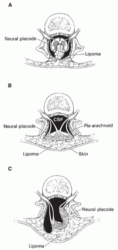
Figure 9-19 Schematic illustrating spinal lipomas and lipomyelomeningoceles. A. Subpial-juxtamedullary lipoma. The spinal cord is open in the midline dorsally with the lipoma situated between the nonapposed lips of the placode. B. Lipomyelocele. This lesion is very similar to a myelocele with two additional characteristics. The lipoma lies dorsal to and is attached to the surface of the placode. This lipoma is continuous with subcutaneous fat. Equally important is the fact that an intact skin layer overlies the lesion, making this an occult spinal dysraphism. C. Lipomyelomeningocele with rotation of the neural placode. When the lipoma is asymmetric, it extends into the spinal canal and causes the ventral meningocele to herniate posteriorly and the dorsal surface of the placode to rotate to the side of the lipoma. This rotation brings the contralateral dorsal root (in this case the right dorsal root) into the midline posteriorly, putting it at increased risk for surgical trauma. Moreover, the left roots are markedly shortened by this rotation, limiting the mobility of the cord and impeding the neurosurgeon from completely untethering it.

Intradural lipomas appear on imaging studies as focal, round to oval, masses that most often lie dorsal to the spinal cord (Figs. 9-20 and 9-21) and may expand the spinal canal (Fig. 9-21). Lipomas have a very low attenuation on CT and, therefore, can be detected on studies without intrathecal contrast (Fig. 9-20). They are easily identified on T1-weighted MR images by their lobulated shape and high signal intensity (Fig. 9-21). Their fatty nature can be confirmed, if necessary, by use of fat suppression pulses. MR best identifies compression of the spinal cord.


Figure 9-20 Intradural lipoma CT. A. Axial CT image shows a low-attenuation mass (arrows) filling much of the spinal canal and the interface with the displaced and compressed spinal cord on the left side of the canal. B. Curved sagittal CT reformation of the lumbosacral spine demonstrates the low-attenuation lipoma (black arrows) and multiple osseous anomalies of segmentation (often incorrectly called “fusion”) in the vertebral bodies and posterior elements (white arrows).


Figure 9-21 Intradural lipoma MRI. Sagittal (A) and axial (B) T1-weighted images show the hyperintense lipoma (arrows) dorsal to the compressed spinal cord. The spinal canal is expanded by the mass.


Lipoma with Dorsal Defect (Lipomyeloceles and Lipomyelomeningoceles)


Description and presentation

Lipoma with dorsal defect occurs when a lipoma that is tightly attached to the dorsal surface of a neural placode extends dorsally through a bony spina bifida to be continuous with subcutaneous fat (42,148,161). Terminal lipomas are probably identical to caudal lipomyelocele; these lipomas attach to the cord at the conus medullaris, which is almost invariably low in position due to tethering, and then extend dorsally through a sacral spina bifida. They constitute approximately 20% of skin-covered lumbosacral masses and between 15% and 50% of cases of closed spinal dysraphism (35,162); they account for about 75% to 85% of spinal lipomas (35). Patients are typically female. Affected patients most commonly present with a rather soft lumbosacral mass, less commonly with sensory loss in the sacral dermatomes, bladder dysfunction, lower extremity weakness, orthopedic deformities of the foot, scoliosis, and/or leg pain (148,150,163). When a lumbosacral mass is present, patients typically come to medical attention before the age of 6 months. If the mass is subtle, or no mass is present, clinical presentation is typically the result of neurologic or urologic deficits that are noticed between the ages of 5 and 10 years; patients may occasionally go undetected into adulthood (150,164). As children with lumbosacral masses are usually detected early, 40% to 45% of such children are neurologically normal on initial examination (42,148,161). It is not clear how many of these asymptomatic patients will eventually become symptomatic, with different series reporting that as few as 16% and as many as 88% of such children eventually develop progressive neurologic symptoms if untreated (148,150,163,164). There is no debate that most symptomatic patients will have progression of symptoms if untreated (148,150,163,164).

Lipomas with dorsal defect usually occur in the lumbosacral region of the cord and tether the cord at that level. Anatomically, lipomyelomeningoceles and lipomyeloceles are very similar to myelomeningoceles and myeloceles, respectively, with two important additional characteristics: (a) a lipoma is attached to the dorsal surface of the placode, and (b) an intact skin layer overlies the lesion (closed spinal dysraphism).


Imaging characteristics

Patients with lipomyeloceles have a normal-sized subarachnoid space ventral to the placode; the cord and the junction between the placode and the lipoma, therefore, are within the spinal canal (Figs. 9-22 and 9-23). The lipoma extends dorsally through the spina bifida and is continuous with subcutaneous fat. The focus of continuity with extraspinal fat is sometimes quite small (Fig. 9-22) but is always present. The lipoma appears as an echogenic mass on ultrasound, as a mass of very low attenuation on CT, and as a mass with short T1 and T2 relaxation times on MR. The spinal cord can have a number of different shapes depending upon the morphology of the lipoma. It is usually crescentshaped, arching over the ventral surface of the lipoma. However, if the intracanalicular portion of the lipoma extends laterally on both sides of the cord, the placode may appear pointed, with the tip of the point directed posteriorly between the lateral extensions of fat. Rarely, wellformed bones (Fig. 9-22) or hamartomatous masses (165) are found in the lipoma.

Only gold members can continue reading. Log In or Register to continue

Stay updated, free articles. Join our Telegram channel

Nov 11, 2018 | Posted by in NEUROLOGICAL IMAGING | Comments Off on Congenital Anomalies of the Spine

Full access? Get Clinical Tree

Get Clinical Tree app for offline access