Hydrocephalus


Hydrocephalus


Charles Raybaud


Introduction

Hydrocephalus is one mechanical complication of many different diseases. It may be defined as a process in which the cerebrospinal fluid (CSF) compartment is actively enlarged at the expense of the brain tissue. It may develop at any age and is common in all age groups, but especially so in children. The basic nature of hydrocephalus is relatively easy to understand: as the ventricles actively increase in size, which results in a progressive compression of the brain tissue, some CSF must be diverted in order to avoid damage to the brain. Establishing the diagnosis also is relatively simple (in most cases): increased ventricular size, effacement of the subarachnoid spaces, macrocephaly, and, typically, an obstruction somewhere along the CSF pathway; common examples include midline tumors, aqueductal stenosis (AS), and arachnoid loculations. It is also important to remember that hydrocephalus is a “mechanical” disease, and as such, it may be a consequence of many different brain diseases (neoplasia, malformation, infection, hemorrhages, etc.), which make its evaluation more complex in individual patients.

Obstructive hydrocephalus is still commonly understood on the basis of concepts formulated early in the last century by Dandy and Blackfan (1): CSF is produced by the ventricular choroid plexuses and absorbed by the peripheral meninges; its transventricular and extracerebral circulation results from a corresponding arteriovenous pressure gradient; an obstruction anywhere along this path can cause CSF accumulation upstream. Communicating hydrocephalus is a subset of the condition in which no ventricular obstruction is found. This concept implies that for the CSF to circulate in normal individuals, the intraventricular pressure must remain higher than the extracerebral pressure, and this model is not really corroborated by facts. Pericerebral spaces are maintained in normal subjects, as is demonstrated by MR imaging; in addition, the normal tuber cinereum is convex upward. Both of these features indicate that the intraventricular pressure is not higher than the cisternal pressure. An “accumulation” of fluid preventing its peripheral absorption would be expected to result in progressively increasing intracranial pressure (ICP), but pressure may remain normal for months (or years) in patients with chronic hydrocephalus. The presence of stable ventriculomegaly with isolated AS proves that CSF may be absorbed within the ventricles. Indeed, clinical practice demonstrates that different pathophysiologic processes may result in different subtypes of hydrocephalus of different degrees of severity, with different radiologic pictures, different therapeutic approaches, and different prognoses (2).

In the decades following the seminal concepts of Dandy and Blackfan, new facts have emerged. In the 1950s, Bering demonstrated that heavy water could exchange freely between the CSF spaces, the parenchyma, and the blood (3,4), implying that the parenchyma can easily contribute to both the production and the absorption of CSF. His later experimental studies of obstructive hydrocephalus without or with choroid plexectomy suggested that the ventricular enlargement could be explained by the loss of the normal dampening of the systolic pressure wave (5,6). This was better demonstrated by further experiments of Pettorossi and Di Rocco (7,8,9). In the 1990s, the advent of MR imaging made noninvasive studies of the CSF dynamics possible in normal human subjects: it was then demonstrated that the force upon CSF resulting from arterial pulsations is much stronger, and displaces much larger CSF volumes, than the mean arteriovenous gradient (10,11,12). In the last two decades, the identification of the aquaporin (AQP)-mediated water channels in the choroid epithelium (AQP1) (13), the ependymal cells (AQP4), and the perivascular, subependymal, and subpial astrocytic end feet (AQP4) (14) provided cytologic support to the concept that water can easily cross any interface between the CSF spaces, the parenchyma, and the vessels.

The first part of this chapter will review the anatomy and development of the CSF spaces, the processes involved in the production and absorption of the CSF, the forces driving the movements of the CSF, and how these factors may mechanically result in hydrocephalus. The second part will address the diagnostic issues of hydrocephalus: clinical presentation, imaging, and diagnostic features, including a review of the specific subtypes relating to different etiopathogenic factors or contexts. The last part will deal with the treatments of hydrocephalus: the surgical techniques, the results, and the complications.


CSF and CSF Spaces

Classically, the total CSF volume has been estimated to be 50 mL in newborns, 60 to 100 mL in children (15), and approximately 150 mL in adults; ventricular volume is small in infants (10-15 mL) and increases progressively in children (16) up to a ventricular volume of 25 mL in adults. Modern studies using volumetric MR imaging in adults have found higher values: 270 mL for total CSF volume (about 150 mL for the intracranial spaces and 100-120 mL for the spinal spaces (17)) or
even 230 to 270 mL for the intracranial spaces (18). This is an important concept because it implies that, just by pushing the brain to fill the pericerebral spaces, the ventricular volume may increase from 25 to 150 mL and more without changing the brain volume (a mathematical simulation of this process yields similar results) (19). The CSF is composed of water (99%), ions (Na+, K+, Ca++, Mg++, Cl, HCO3), proteins, amino acids, creatinine, urea, lactate, phosphate, and glucose (20,21); protein concentration is higher in newborns (especially those born preterm) than in older children, while glucose concentration is slightly lower (22). CSF serves as a transport vehicle for movement of various neurotrophic substances, signaling molecules, neurotransmitters, and other metabolites, to and from the brain. The concentration of these substances in the CSF is “analyzed” throughout life by the circumventricular organs, portions of the ventricular walls that are devoid of CSF-brain and blood-brain barriers and, thereby, allow the hypothalamus to react in response to changing CSF composition (23,24).


Anatomy


The Container: Skull and Spine

CSF and interstitial fluid (ISF) together form the extracellular fluid compartment of the central nervous system (CNS). CSF is contained within the ventricles and arachnoid spaces and surrounds the brain and spinal cord. It acts as a transportation medium as well as a cushion for the neural tissue, protecting it from mechanical damage. The immersion of the brain reduces its weight substantially (˜97%), hence lessening the effects of both intracranial and extracranial forces.

The skull is composed of the vault and the base. Although the base is traversed by many nervous and vascular foramina, the mature skull may be considered a closed box, except for the foramen magnum that opens into the spinal canal. The dura that lines the inner calvarial table forms folds that project into the cranial cavity to form the falx cerebri in the sagittal plane, and the tentorium cerebelli between the brain and the cerebellum. Between the insertions of the falx and tentorium on the calvarial dura run the dural superior sagittal, transverse, and sigmoid venous sinuses, which themselves are lined with arachnoid villi. Blood is conveyed into the internal jugular veins through the jugular foramina when the body is supine, but through numerous other veins and dural venous channels, into the neck and the spinal venous system when the body is erect (25). Within the insertion of the falx on the tentorium, the triangular-shaped straight sinus conveys the venous blood of the vein of Galen into the torcular Herophili and the transverse sinuses. On either side of the sella turcica, the complex plexular cavernous sinuses drain the orbital and sylvian veins toward the pterygoid plexuses and petrosal sinuses. While the mature skull is rigid, the fetal and infantile skulls are more plastic due to the fibrocartilaginous sutures and fontanelles. In volume, relative to the spinal canal, the cranial cavity is also much larger in fetuses and infants than in adults.

The spinal canal is enclosed by the vertebral bodies, the neural arches and ligaments from the foramen magnum to the sacral segment. Unlike in the skull, the dura is not attached to the bone but separated from it by the epidural fat and epidural veins. It also forms 30 pairs of nerve sheaths around the 30 pairs of emerging spinal nerves in the intervertebral foramina. This anatomy confers enough elasticity to the meningeal sac to accommodate the pulsatile craniospinal influx of CSF at each cardiac stroke.


The CSF Spaces


The ventricles

The ventricular system comprises the lateral, third, and fourth ventricles. The foramina of Monro lead from the lateral to the third ventricle. The mesencephalic aqueduct of Sylvius, connecting the third and fourth ventricles, is the narrowest portion of the ventricular system. The frontal horn, body, and atrium of the lateral ventricle circumscribe the dorsal and posterior aspects of the caudate and thalamus. They are limited cranially by the fibers of the corpus callosum; laterally by the caudate nucleus and the hemispheric white matter; medially by the septum pellucidum, hippocampal commissure, and fimbria; and from the foramen of Monro to the temporal uncus by the choroid fissure, which contains the anterior and the posterolateral choroid arteries. The frontal horn and body of the ventricle normally are narrow, roughly triangular with one acute lateral angle. The occipital horn is virtual in normal children, surrounded by white matter, with the calcar avis (primary visual cortex) lining it medially. The temporal horn is surrounded by the hippocampus medially, the parahippocampal white matter inferiorly, and the deep temporal white matter superiorly. The thinnest part of the ventricular walls is the medial wall of the atrium, which may expand medially and form a diverticulum in case of chronic hydrocephalus. The walls of the lateral ventricles contain the subependymal veins, which are directly exposed to the high intraventricular pressure in patients with hydrocephalus.

The midline, thin third ventricle is limited anteriorly by the hypothalamus and posterolaterally by the thalami (with their midline adhesion, the massa intermedia). The anterior part of its floor is the tuber cinereum, which is normally convex upward, and the posterior part is the cap of the midbrain. The anterior wall is the lamina terminalis limited by the optic chiasm inferiorly and the anterior commissure superiorly. The supraoptic and infundibular recesses are located above and behind the optic chiasm, respectively. The lamina terminalis, tuber cinereum, and anterior-inferior recesses may dilate and bulge significantly in case of obstructive hydrocephalus. The aditus of the aqueduct is between the cap of the midbrain and the posterior commissure, below the pineal recess (at the base of the pineal gland), which itself is below the (variable) suprapineal recess; it is lined by the subcommissural organ (SCO). The roof of the third ventricle is the tela choroidea and its plexus, which are continuous laterally with the choroid fissure of the body of the lateral ventricles. It contains the internal cerebral veins and the posteromedial choroid arteries. The third ventricle contains three circumventricular organs: the vascular organ of the lamina terminalis anteriorly and the lateral subfornical organ superiorly, in addition to the SCO posteriorly.

The cerebral aqueduct curves gently caudally through the midbrain from the third to the fourth ventricle. It measures about 15 mm in length in adults, with two narrowings corresponding to the anterior and posterior colliculi and an ampulla in between; its diameter varies from 0.5 to 2.8 mm (26). It lies caudal and ventral to the tectal plate and is surrounded rostroventrally by the gray matter of the oculomotor nuclei.

The fourth ventricle lies on the dorsal aspect of the brainstem. It has a rhomboid shape with a rostral half that is pontine and a caudal half that is medullary. The rostral half is limited laterally by the anterior, middle, and inferior cerebellar peduncles, the caudal half by the lower rhombic lips. The vermis forms the roof of the rostral half, and the vermian nodulus encroaches caudally upon the ventricular lumen, giving it a triangular shape on a midline sagittal plane; the dorsal apex of the triangle is the fastigium. The lateral angles form the lateral recesses. The caudal half of the ventricle opens into the cisterna magna. Developmentally, this opening results from the dorsal expansion of the embryonic tela choroidea (Blake pouch) into the infravermian space; this tela choroidea therefore lines the inferior aspect of the vermis and cerebellar hemispheres and attaches to the posterior occipital dura, so limiting the cisterna magna, which itself communicates with the posterior fossa and spinal cisterns, but not with the vermian cisterns: the cisterna magna is truly an expansion of the fourth ventricle and as such
does not contain arachnoid septations (27). The dorsal attachment of the tela choroidea to the dura forms a transverse membrane that separates the cisterna magna from the retrovermian cistern (28). Conventionally, the dorsal aperture is called the foramen of Magendie and the openings of the lateral recesses into the cerebellopontine angles, the foramina of Luschka. The choroid plexus is attached transversely between the lateral recesses, along the inferior medullary velum: it is actually located in the cisterna magna as much as in the fourth ventricle. The caudal apex of the fourth ventricle is continuous with the (usually virtual) central canal of the caudal medulla and spinal cord and is covered by the obex; this is where the area postrema (most caudal circumventricular organs) lies.


The extracerebral spaces

The pericerebral CSF spaces of the cerebral convexity are intra-arachnoid, contained between the deep layers of the dura and the cortical pia. They are wide in fetuses, so that the cortex does not abut the calvarium. They become smaller in normal infant and become hardly visible during childhood and adolescence along the temporal lobes (where the gyri become imprinted on the inner table and form the cerebriform markings), while usually remaining apparent over the upper frontoparietal convexities. The cisterns are widened portions of the arachnoid spaces interposed between the hemispheres or between the brain and the base of the skull. They are named according to their location. They are typically traversed by crossing nerves, arteries, and veins, and except for the cisterna magna (an expansion of the fourth ventricle), they contain arachnoid septations that may become inflamed and fibrotic and prevent the free circulation of CSF. In the spine, the CSF spaces surround the cord and cauda equina and may be considered a single cistern. Rostrally, they communicate widely with the cisterna magna and the perimedullary cistern through the foramen magnum. In the spine, they surround the cord and are crossed by the anterior motor and posterior sensory nerve roots (which form the cauda equina in the lumbosacral segment) with their accompanying arteries and veins. They contain 21 pairs of arachnoid dentate (or denticulate) “ligaments” that attach the cord laterally, the septum posticum that attach the cord posteromedially, and the filum terminale. Laterally, the arachnoid spaces extend into the nerve sheaths down to the sensory ganglions. Besides providing expandability to the meninges and allowing the sliding motion to the spinal nerves, the nerve sheaths seem to be significant CSF absorption sites. Lateral encystments of the sheaths may be seen at the sacral level and are called the cysts of Tarlov.


Vessels and choroid plexuses

The cerebral arteries originate from the internal carotid and the vertebral arteries. They are organized with multiple levels of anastomosis (basilar artery and circle of Willis in the cisterns, pial anastomoses on the surface). From the pial network, numerous perforators enter the brain from the periphery toward the ventricular wall. The only (apparent) exception are the choroid arteries that supply the intraventricular choroid plexuses and deep subcortical gray matter. Arterioles are much more numerous in the gray matter, especially the cortex, than in the white matter, resolving into a capillary bed that perfuses all cortical layers. The cerebral blood flow is extremely high (CBF = 50 mL/100 g/min in the adult, much more in infants and children), whereas the cerebral blood volume is small (CBV = 4 mL/100 g of tissue). As both CSF and parenchyma are incompressible, the capillary bed is the “weak,” vulnerable compartment when the ICP is increased.

The cerebral veins are organized in two drainage systems. One, superficial, drains the cortex and the subcortical white matter toward the pial cortical veins; these join into the bridging veins that cross the CSF spaces toward the dural venous sinuses. The other, deep medullary, drains the deep white matter and part of the central gray matter and forms the tributaries of the subependymal veins, which course in the ventricular walls toward the internal cerebral veins, the vein of Galen, and the straight sinus.

The choroid plexuses are attached along the choroid fissures of the lateral ventricles (forming a bulky glomus in the atria), in the roof of the third ventricle and along the inferior medullary velum of the fourth ventricle. They secrete the CSF. In the early, still poorly vascularized fetal brain, they are loaded with glycogen (29). Together with the pulsating vessels and parenchyma, they participate in the dynamics of the CSF. Interestingly, choroid plexuses are able to regrow after having been partially resected.


Development


Choroid Plexuses

The closure of the neural tube occurs by about 28 postconceptional days in the human (see Chapters 5 and 9). Until then, the apical (future ventricular) surface of the neuroepithelium is bathed in the primitive amniotic fluid and fed directly from it. After it closes, the neural tube is surrounded by the meninx primitiva, which itself is invaded by rostral “branches” (only primitive capillaries at this stage) of the developing cardiovascular system, from which nutrients diffuse into the neuroepithelium (29), and fluid produced by the neuroepithelium fills the central lumen (30,31). Rostral segments of the neural tube expand to form the basic pattern of the ventricular system: prosencephalic (forebrain), mesencephalic (midbrain), and rhombencephalic (hindbrain) vesicles. During the second gestational month (5-7 weeks), the midline dorsal meningeal mesenchyme invaginates into the lumen of the fourth, lateral, and third ventricles to form the choroid plexuses of the ventricular system, while the prosencephalon divides dorsally and forms the cerebral hemispheres. The water channel AQP1, which transports water across the plexular epithelium, is expressed from the very early stages of plexular development in the rat fetus (32) and as early as the seventh week in the human fetus, first in the tela choroidea of the fourth ventricle and then, 1 week later, in all choroid plexuses (33). Enzymes used for CSF secretion have been demonstrated in a 9-week human fetus (34). Choroid plexuses are composed of a secretary cuboidal epithelium limiting a stromal core of capillaries and connective tissue (choroid interstitium) (21). The vessels present a fenestrated endothelium allowing water and small molecules to egress into the stroma. Joined by tight junctions, the epithelial cells form the blood-CSF barrier and act as active transporters (for water, ions, proteins, nutrients, and neurotrophic factors) from the plexular stroma to the CSF. Microvilli on their apical surface increase the surface area of the choroid plexuses up to 200 cm2 in humans (21). Cells of the immune system (macrophages, dendritic cells, Kolmer epiplexus) are associated with the plexus, sometimes being transported through them as the plexus does not have a blood-brain barrier (21,35). Microglia from the choroid plexus colonize the CSF and subsequently invade the deep cerebral parenchyma as early as 5.5 weeks (21,36). The choroid plexuses deliver, and the ventricular CSF carries oxygen, glycogen, proteins, and growth and neurotrophic factors to the periventricular germinative zone (21,31,37,38). This function of the choroid plexuses persists in later gestation as a significant complement to parenchymal vessels and remains important in postnatal life (31,39,40). The choroid plexuses may also remove endogenous and exogenous substances from the CSF (41,42). Their importance is reflected by their size: they fill approximately 75% of the ventricular lumen during the third month of gestation and reach their largest size about 26 to 28 weeks. It has been suggested that they
might play a role in the expansion of the cerebral ventricles by maintaining an optimal hydrostatic pressure (43). This could also result from their arterial pulsatile force (see below). Although histologically similar, the choroid plexuses present different and specific molecular secretion patterns (“secretome”) in the lateral, as compared with the fourth, ventricles that are consistent with correspondingly specific forebrain and hindbrain receptors (21).


Ependyma

For most of the first two trimesters, the ventricles are lined with the undifferentiated germinative ventricular epithelium. True ependyma differentiates from the radial glia as the ventricular zone (“germinal matrix”) regresses, to form a continuous lining at about 26 to 28 weeks (44,45). In adults, large areas of it disappear (45). The water channel AQP4 is expressed along the ventricular lining of the hippocampus after 14 weeks and sometime before 22 to 23 weeks in the ependymal and subpial layers of the neocortex: the expression being strongest at about 28 weeks, when the ependyma is complete (46).

Ependymal cells are ciliated cuboidal cells, each with an average of 16 motile cilia measuring approximately 13 µm in length. The coordinated beating (28-40.7 Hz frequency) of this ciliated epithelium is believed to direct neurotrophic factors and guidance molecules from the plexuses toward their targets along the ventricular walls, to “clean” the ventricular wall, notably the recesses, from deposits and to facilitate CSF flow in narrow channels (30,31,45). It has been shown in the mouse embryo that the initial orientation of ciliary beating depends on the direction of the ventricular CSF flow (47). In fetal, but also in more mature, brains, the ependyma supports the progenitor cells of the subventricular zone (48). Vascular endothelial growth factor (VEGF) is expressed in the fetal ependyma during the second half of gestation (45). The ependyma is altered by hydrocephalus (45), although some ependymal repair might occur (49). The ependymal denudation that results from fetal hydrocephalus has been associated with and is felt to be the cause of the development of periventricular nodular heterotopia (see Chapter 5) (50).


Extracerebral Spaces

The extracerebral CSF spaces develop during the second month when the meningeal layers differentiate. The intercellular spaces of the meninx primitiva enlarge progressively and coalesce to form a fluidfilled trabeculated (sub)arachnoid space (51,52), limited peripherally by the arachnoid layers of the dura mater (53) and centrally by the pia that covers the surface of the cortex. It also forms the arachnoid septations and the sheath of the leptomeningeal blood vessels, thereby limiting the perivascular spaces (PVS). The pia also ensheaths the vascular intraparenchymal perforators, thereby forming the Virchow-Robin spaces (VRS), a combination of subpial space and PVS (this will be detailed in the following section of this chapter) (54,55,56,57). The leptomeningeal “liquefaction” process begins ventral to the pons and midbrain at 5 weeks, extends both caudally and rostrally at 6 weeks, develops dorsally to the fourth ventricle at 7 weeks, and becomes generalized (if not completed yet) about 8 weeks (52). This is before the opening of the fourth ventricular outlets, which are not identified in humans before weeks 11 to 12, as a result of the process of expansion and attenuation of the caudal, medullary part of the rhombencephalic roof (Blake pouch) (27,58). In the fetus, the pericerebral spaces (as demonstrated by fetal MR imaging) are prominent until close to term, but they are smaller in premature babies of similar gestational age (2); this likely reflects the decrease of intracranial water associated with the venous pressure drop that occurs at birth when the pulmonary bed opens and the cardiac circulation reorganizes (59). This implies that, in the fetus, the growth of the skull is driven by the CSF pressure rather than by the growth of the brain: the brain expands within the CSF without being constrained by the cranial vault. This is likely true during childhood as well. High venous pressure (and low arterial pressure) means that the fetal intracranial arteriovenous gradient is less than that of the (postnatal) infant. The vascular pulsatility also is less, as the CBF is assumed to be much lower in fetuses than it is after birth (59,60); at this stage, it may be more prominent in the plexuses than in or around the parenchyma.


Arachnoid Villi

It is generally assumed that fetuses and infants have no functional arachnoid villi, which progressively appear during childhood (socalled immaturity of the absorption sites). However, although no apparent arachnoid villi are found in fetuses, depressions in the dural sinus walls have been described as early as 26 weeks; although these do not become true villi until much later, they may already be functional (61). Other absorption sites such as the nasal lymphatics under the cribriform plates seem to be functional very early.


Secretion and Absorption of the CSF


CSF Production

The daily production of CSF is commonly estimated to be 500 to 600 mL in the adult, with a daily turnover of 3 to 4. This is large when compared to the ventricular size, but it corresponds to a production rate (hence an absorption rate) of 0.35 mL/min, which is too small to be reliably measured by magnetic resonance imaging (MRI)-based CSF flow studies in individual patients. This is an average rate that seems to be fairly constant between individuals and between normal and hydrocephalic patients (62). But the instantaneous rate varies widely during the day, from 0.05 to 0.78 mL/min in adults with nonobstructive hydrocephalus, being higher in the second part of the night, and from 0.25 to 0.40 mL/min in hydrocephalic 5- to 13-year-old children (lowest at 0.25 mL/min, in younger 1-month- to 8-year-old patients), with variations due to physiologic activities such as crying, coughing, or REM sleep (62). It has been shown to vary by a factor of 10 over a 1-hour period, even though the average remains within previously established normal limits (63). As development proceeds, the production increases in proportion to the size of the brain; this would account for differences between males and females (62).


Choroid plexus secretion

Choroid plexuses are still assumed to be the main source of CSF, with estimates ranging from 60% to 90% of the total CSF, the remaining 10% to 40% being produced by the brain and cord parenchyma. Choroid plexuses secrete the CSF in two steps: first, a passive exudation across the fenestrated endothelium into the plexular stroma and then a regulated transport of water, ions, and larger molecules across the epithelial layer into the ventricle (21,64). Within limits, the rate of active secretion appears to be relatively insensitive to osmotic pressure changes (65). The passive exudation in the plexular stroma decreases when ICP increases (64) but obviously never stops completely (the arterial pressure is superior to the ICP as long as a blood flow is maintained); it is regulated by vasoactive substances (norepinephrine, angiotensin II, and serotonin). The active secretion uses ATP hydrolysis to generate a unidirectional flux of ions across the plexus epithelium (41), regulated by neuropeptides like choroidal arginine vasopressin (AVP) and atrial natriuretic peptide (ANP) (64). Water is transported by the cellular water transporter AQP1, which is found in high concentration at the apical side of the choroid epithelial cells (65,66,67). Nutrients, proteins, metabolic precursors, and neurotrophic factors also are secreted
or produced by the epithelium; this “secretome” is both regionalized (different in the forebrain and the hindbrain, adapted to specific receptors in both locations) and evolutive according to the stage of brain development (21).


Contribution of the brain parenchyma

Choroid plexectomy does not result in a lack of CSF (68). Water molecules exchange diffusely across the brain surfaces, as demonstrated long ago by the use of heavy water molecules (3,4,69). In the case of an osmolar shift, this exchange may represent a significant contribution to the total volume of CSF (70). In obstructive tetraventricular hydrocephalus, the extracerebral CSF necessarily is produced from the parenchymal ISF by the surface of the brain and cord (including the surface of the VRS). ISF and CSF together form a single brain extracellular fluid compartment, and they share a similar composition (20). The ISF volume of the CNS is estimated at 100 to 300 mL (41), of the same order of magnitude as the CSF. The water channel AQP4 is densely expressed in the ependyma and astrocytic end feet, which are located in the subependymal layer, around the parenchymal capillaries, and along the subpial glia limitans of the brain VRS surfaces. AQP4 may transport water either way depending on pressure and osmolarity gradients (65,66,67).


The Virchow-Robin spaces

The VRS form blind channels that originate in the depth of the parenchyma and open at its surface, a pattern reminiscent of that of the lymphatic channels, suggesting that they may participate in the drainage of the parenchymal ISF into the arachnoid spaces. They develop from the surface of the brain together with the perforant vessels. The pia is separated from the cortex by the subpial space, and the arachnoid is separated from the leptomeningeal vessels by the PVS (56). When the vessels enter the parenchyma toward the periventricular germinative zone, so forming the perforant arteries and veins (week 8 and after) (70,71,72), they “carry” the leptomeninges with them (55) so that each perforant vessel is surrounded with a PVS, a pial sleeve, and a peripheral subpial space limited by the AQP4-rich glia limitans, which together form the VRS (56). Venous VRS are simpler than the arterial VRS, with a partially attenuated pial sleeve (56). In contrast, VRS are more complex around the basal ganglia perforators, with a double leptomeningeal sleeve (57). VRS do not extend along the capillaries (55,56).

PVS, VRS, and subpial space seem to communicate freely (54). Whether they remain separated from the arachnoid space and convey fluid toward extradural lymphatics (54) or whether the leptomeningeal sheath is fenestrated and allows the fluid to drain into the arachnoid space itself is not known with certainty (56,73,74). The latter, however, seems most likely, as pericerebral CSF is produced in the case of a ventricular obstruction, and no possible alternative structure has been identified that could do it. Recent reports have suggested that arterial and venous VRS together would actually have different roles, the pulsating wave in the arterial VRS driving water toward the parenchyma and the venous VRS draining it toward the arachnoid space (75,76,77). The term “glymphatics” (for glial lymphatic) has been proposed to describe this arrangement (76). However, this concept is not supported by other studies and is still controversial (20,73). It may also be argued that, in the absence of a valve at the opening of the arterial VRS, pulsation may facilitate diffusion but not create a pressure gradient to convey water inward. At this point, it seems most likely that water is transported by the AQP4 channels from the ISF to the VRS and from there to the arachnoid space. This parenchymal contribution might represent as much as 30% of the CSF (41), or even more (12).


CSF Absorption

Obviously, the absorption rate of the CSF in a normal situation equals its production rate: 0.35 mL/min or 500 to 600 mL a day. One study using intrathecal infusion in adult subjects found that absorption does not occur at pressures of 68 mm H2O or below and that it increases linearly with an increasing ICP to reach about 1.5 mL/min at a pressure of 250 mm H2O; it was not measured above that level (15) (the normal range of ICP in the supine position is 100-180 mm H2O or 8-15 mm Hg). Therefore, the normal absorption capacity of the system is ample as long as the absorption mechanisms are intact. These mechanisms are described below.


Transdural absorption

CSF absorption is known to occur across the arachnoid villi (forming Pacchionian granulations), in which arachnoid tissue protrudes into the lumen of the dural venous sinuses across their wall. Although fetuses and infants have no or only few apparent arachnoid villi, depressions in the dural sinus walls have been described as early as 26 weeks, becoming true villi only much later (61). Anatomically, the villi appear to be oneway valves, which open at a pressure difference of 20 to 50 mm of water (61); drainage depends on the presence of a CSF-venous sinus hydrostatic pressure gradient (some evidence suggests that absorption may be by pinocytosis instead) (61,78). In addition to the villi, other transdural drainage routes exist: dural clefts along the parasagittal dura (different from the classic villi) (79); venous plexuses of the cavernous sinuses or around the internal carotid artery and pituitary gland in the sellar region (80); olfactory nerve fibers across the cribriform plate toward the nasal mucosa and lymphatics (81); optic nerves sheaths toward the orbital lymphatics (82,83); and other cranial and spinal nerve outlets with their own arachnoid villi (61,84). It is not known whether these different pathways are hierarchical, depend on age and maturation (61,84,85), or are simply synergetic, but all contribute to the CSF absorption. Importantly, all are “passive” routes dependent on a CSF-peridural pressure gradient only.


Parenchymal absorption

Although absorption of the CSF by the brain has been denied on the basis of tracer studies, it has been shown that water (labeled with deuterium) would diffuse easily from the ventricles to the parenchyma and, from there, to the blood (3,4,22). This view was strengthened in the last two decades by the identification of the aquaporins water channels AQP1 and AQP4, which transport water only—not ions, and not tracers other than water labeled with hydrogen isotopes deuterium or tritium (13,14,20,32,33,46,65,66,67,73). AQP4 is strongly expressed in the ependymal cells and subependymal astrocytic end feet of the ventricular walls. The transport of water between CSF and ISF may occur in both directions, according to pressure and osmolarity gradients; the ventricular walls may adjust to specific contexts by removing (or providing) water from (or to) the ventricular CSF. In animal experiments, it was found that acute occlusion of the aqueduct does not modify the intraventricular pressure and produces no ventricular dilation over a 2-hour period, supporting the hypothesis that most of the CSF produced by the choroid plexus during that time was absorbed by the ventricular surface (86,87). The same group showed that there is no outflow of CSF from a cannulated aqueduct when CSF pressure is normal (70). Although such experiments may be criticized for their nonphysiologic conditions, the results and conclusions are consistent with the clinical example of chronic untreated ventricular obstructive hydrocephalus: water (not necessarily “complete” CSF with its solutes) is clearly absorbed by the ependyma. As the pericerebral CSF is obviously produced by the brain surface/VRS as well, ventricular water may be transported to the ISF and, from there, to the parenchymal capillaries as well as to the arachnoid spaces across the brain surface and VRS.


CSF Dynamics and Related Disorders

Several types of CSF flow may occur in the CNS. Bulk flow is the net movement of a volume of fluid between two points. Oscillatory flow describes a to-and-fro displacement of a volume of fluid between two points. Diffusion describes the motion of individual molecules within a fluid; it may occur in a volume of fluid that is otherwise static. Within the cerebral ventricles, the term cilia-directed near-wall flow describes the movement of the thin layer of juxtaependymal CSF that is displaced by the coordinated beating of the ependymal cilia.


Vertical Bulk Flow Versus Transverse Bulk Flows

As discussed above, the translation of water molecules from the secretion sites to the absorption sites is referred to as a bulk flow. It is traditionally held that this translation is from the choroid plexuses to the dural sinuses along ventricles and cisterns. However, theoretical (massive expression of AQP4), clinical (untreated chronic obstructive hydrocephalus), and experimental (see above) evidence strongly suggests that a significant volume or even the whole amount of the CSF secreted by the plexuses may be absorbed by the ventricular walls, even in normal conditions. If CSF production and CSF absorption sites are multiple, it is possible to conceive that multiple, separate “segmental” bulk flows may simultaneously coexist. This concept is consistent with the recent finding that choroid plexuses in either the lateral or the fourth ventricles secrete trophic factors that selectively target the cerebral hemispheres or the hindbrain (21), which is possible only if these factors are not dispersed by a global bulk flow. Indeed, evidence now suggests that there are several “transverse” bulk flows (in the lateral ventricles, the third ventricles, the fourth ventricle, cranial extracerebral spaces, spinal canal), rather than a single “longitudinal” bulk flow from the plexuses to the periphery (Fig. 8-1). This doesn’t, however, mean that such hydrodynamic segments are isolated; exchanges almost certainly occur along the way, maintained by molecular diffusion and by mixing due to the CSF pulsations. More importantly from a point of view of hydrodynamics, the lack of a physical separation prevents a transmantle pressure gradient from occurring if a secretion-absorption disequilibrium were to develop between isolated CSF compartments. Above all, the freedom of to-and-fro CSF motion allows the force created by the arterial systolic wave to be dampened by the viscoelastic response (compliance) of the system.


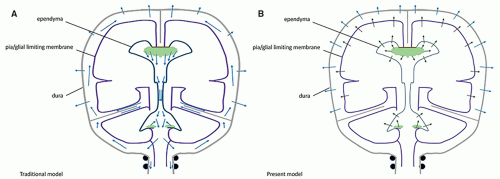
Figure 8-1 Models of CSF circulation: bulk flows. A. Traditional model (vertical bulk flow). This model postulates that the CSF, secreted by the choroid plexuses, would flow along the ventricles, then into the cisterns, and then toward the convexity to be absorbed at the level of the arachnoid villi. B. Modern model (transverse bulk flows). The new models stipulate that the CSF secreted in the lateral, third, and fourth ventricles is absorbed locally at the corresponding ependymal levels, allowing specific signaling molecules to be secreted and reach their local subependymal targets at each level. Similarly, the pericerebral CSF produced by the brain surface is absorbed transdurally at the periphery.


CSF production and absorption mismatch

Dandy and Blackfan explained “communicating” (nonobstructive) hydrocephalus by a poor absorption as opposed to an obstruction and implicated inflammatory leptomeningeal changes (1). As they assumed that absorption occurred diffusely in the leptomeninges, it is not clear whether they meant a true absorption disorder or a cisternal obstruction. This uncertainty persists today (88). Abnormal absorption was considered to be the mechanism of the “secondary” NPH, as a late complication of a subarachnoid hemorrhage, an infection, or a trauma: the intrathecal infusion test was intended to evaluate the CSF absorption capacity, while the ventricular pooling of contrast in isotopic or CT cisternographies was assumed to reflect a poor peripheral absorption. Yet this “absorptive” model seems to correlates poorly with the results of CSF shunting (89), and the very concept of nonobstructive hydrocephalus as an absorption disorder is now debated (90). A recent classification of hydrocephalus suggests that most cases of “communicating hydrocephalus” are due to a hidden obstruction (91). However, there is clinical evidence that restriction to the cerebral venous outflow may result in an accumulation of CSF and that hydrocephalus develops in the case of an excessive hypersecretion of CSF (e.g., choroid plexus papilloma) although there is no evidence of a CSF pathway obstruction.


Pulsating Flow and Cerebral/Thecal Compliance

If ventricular CSF absorption is maintained and cannot explain hydrocephalus associated with a ventricular obstruction, what is the origin of the force that dilates the ventricles? After O’Connell in 1943 (92), Bering addressed this question in two reports (5,6). Asserting that the “secretion pressure” is unable to produce the ventricular dilation, he stipulated that “each pulsation of the choroid plexuses sets up a pressure gradient which tends to force CSF out of the ventricles. This acts as an unvalved pump, imparting a to-and-fro motion to the CSF” (5) and that “[The] compression wave in the cerebral ventricles […] normally is absorbed in part by the brain, in part by pumping of the CSF out of the ventricles and finally by the veins […]. When either or both pathways
of CSF or venous drainage […] are blocked, the intraventricular pulse is increased correspondingly and this entire force must be absorbed by the brain” (6). This dual dampening process constitutes the compliance of the system. The loss of elasticity results in less attenuation of the force of the pressure wave, so that this force is fully exerted against the brain tissue. The dampening effect is also lost if the venous pressure is increased; again, the force of the pressure wave is exerted against the parenchyma. In both instances, a progressive loss of brain tissue may ensue. Modern studies have shown that the pulsatile force originates not from the plexuses only, but from the whole intracranial vascular system (pericerebral, intracerebral, and choroid) (8,12,69,93,94). They have emphasized its importance in the CSF hydrodynamics (7,9,11,93,95,96,97,98,99,100,101), and MR imaging has become a major investigation tool in this regard (10,93,94,99,101). Most of the force of the systolic inflow is buffered by displacing blood along the vascular system (94%); capillary and venous resistance therefore are potentially major factors of hydrocephalus (Table 8-1). Significantly smaller oscillatory volumes of CSF are displaced by the residual vascular expansion out of the ventricles and out of the cranium toward the more elastic spinal dural sac; in normal conditions, they have been evaluated at 1.7 mL/min at the aqueduct (with peak velocities of 10 cm/s, however) (10,11,102), 14.5 mL/min at the incisura, and 39 mL/min in the upper cervical canal (10) (Fig. 8-2). These movements produce the flow void artifacts that are easily detected on T2SE WI (103). In comparison, the amount of fluid displaced through the absorption sites is negligible: 0.35 mL/min, which is a hundred times less than the CSF volume displaced at C2-C3 (Table 8-1; Fig. 8-2). The prominent flow voids in the partially enclosed prepontine, suprasellar, and sylvian cisterns (104,105) also likely reflect local turbulences, probably due to basilar and carotid/sylvian arterial pulsations. The lack of an aqueductal flow void in normal neonates (personal data) can be explained by the elastic sutures/fontanels allowing the pulsating force to be fully centrifugal.


Table 8-1 Relationships of Blood and CSF Flows


Volumes


Ratio


Cerebral blood flow 50 mL/100 g/min, brain 1300 g


650 mL/min


100%


CSF displaced


Total (C2-C3)


39 mL/min


6%


Supratentorial (tentorial incisura)


14.3 mL/min


2.23%


Aqueduct


1.7 mL/min


0.26%


CSF absorbed


0.35 mL/min


0.05%


Blood displaced


611 mL/min


94%


Data partly from Enzmann DR, Pelc NJ. Cerebrospinal fluid flow measured by phase-contrast cine MR. AJNR Am J Neuroradiol 1993;14:1301-1307.


Consequences of a compliance decrease

The loss of compliance is an essential factor of every type of hydrocephalus. It may result from increased impedance to CSF displacement or by restriction of the dampening space. Bering and Sato suggested that it could be explained by the physics of the interaction of the arterial pressure wave with the viscoelastic brain and theca (106). It has been shown that the systolic pressure wave presents three peaks P1, P2, and P3, such as P1 > P2 > P3 (96,97,98,99,107,108). P1 corresponds to the systolic inflow, P2 to the viscoelastic response of the vessels, ventricular pathway and theca, and P3 to the venous systolic wave (as the brain circulation is systolic-diastolic) (Fig. 8-3A). If the system is compliant, the shape of the curve is preserved, and the amplitudes of the three peaks are the same inside the ventricles and outside the brain. If the system is not compliant, for example, because the ventricles are obstructed, P2 inside the ventricles becomes as high as or higher than P1 (Fig. 8-3B), while it remains unaffected in the extracerebral CSF spaces: a transmantle force gradient occurs at each systolic pulse that displaces the mantle toward the pericerebral space. However, while this may explain how the pulsation wave dilates the obstructed ventricle at each systole, it does not explain how this dilatation may be maintained or may increase: as the CSF production is unlikely to be increased, it is the absorption that must be decreased. This may be explained by the same physical process. The increased P2 peak being higher than P1 and P3 (P2 > P1 > P3) compresses the capillary bed and veins and, by compromising the appropriate ISF drainage, compromises the transependymal absorption. As a normal absorption would have been, at most, a third of a milliliter per minute, a partial decrease does not amount to much, and only minute amounts of fluid are accumulated in the ventricles at each systole. Ventriculomegaly develops very slowly: in young children, this gives the skull time to expand, so that the brain volume may actually remain unchanged until a new balance is reached between the pulsating wave force and the viscoelastic parenchyma. By increasing the ventricular surface, the process may even increase the capacity of absorption as long as the ependyma is not injured. This situation would correspond to the features of the classic chronic “compensated” hydrocephalus. A particular aspect of this model is that because the pericerebral spaces become effaced, the outward pulsatility is prevented, and pulsatility is fully redirected inward. The amplitude of the pulsation wave is therefore increased within the ventricle; if the aqueduct is open (cisternal obstruction, nonobstructive hydrocephalus), the volume of CSF displaced to-and-fro through it is increased, which explains the increased prominence of the flow void on imaging.


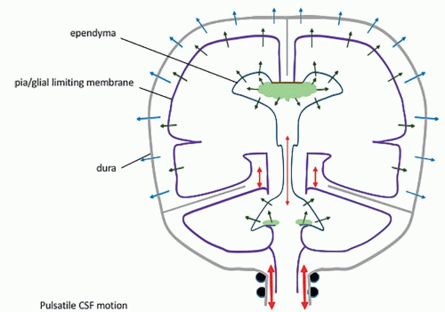
Figure 8-2 Model of CSF circulation: pulsatile flow. The volume of the blood entering the skull at each systole needs to displace a similar volume of fluid. Most represents the systolic-diastolic flow across the capillary bed and into the veins. A small amount of CSF is also displaced toward the more compliant spinal dural sac. The force of the cerebral pulsation is directed outward mostly: the volume of CSF displaced across the tentorium is almost 10 times the volume of CSF displaced across the aqueduct.

This model of ventriculomegaly developing because of a transmantle pulsatile force gradient is sometimes contended because of the absence of any measurable transmantle pressure gradients in patients with communicating or noncommunicating hydrocephalus (109). Such measurements are obviously made at equilibrium (hydrocephalus develops much too slowly to measure the dynamics of the process)
and in the closed cranium. The stability of a mass on the ground does not rule out the force of gravity.


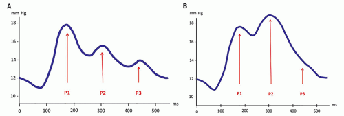
Figure 8-3 The intracranial systolic pressure wave. A. At each systole, the influx of blood into the skull results in a pressure wave. The normal systolic-diastolic pressure wave of the CSF causes three peaks: P1 (systolic pressure), P2 (viscoelastic response of the vessels, parenchyma, and dural sac, i.e., the compliance), and P3 (venous systolic wave). In the normal conditions of compliance, P1 > P2 > P3. B. If the compliance is decreased (the impedance is increased), the shape of the curve is changed with P2 ≥ P1 > P3. This means that in the case of a ventricular obstruction, P2 is higher than P1 within the ventricle, but not outside of it (where the compliance is maintained), and a transmantle pressure gradient occurs at each systole that results in a progressive ventricular dilatation. In addition, by impeding the proper systolic-diastolic circulation through the vascular bed, this abnormal systolic P2 may compromise the venous absorption of ISF and, as a consequence, the transependymal absorption of CSF.


Combined loss of compliance and absorption


Acute hydrocephalus

A common complication in the course of a chronic obstructive hydrocephalus (compensated hydrocephalus) is the development of a high ICP (decompensated hydrocephalus). Several factors have been suggested to explain this: the ventriculomegaly may reach a threshold beyond which absorption cannot adjust anymore; decompensation could be related to the closure of the sutures; or it could be precipitated by a trauma or an infection (110). Pathogenetically, an additional factor needs to be combined with the loss of compliance to explain this decompensation, and a plausible candidate for this is a failure of the absorption processes, such as a progressive increase of the capillary/venous pressure, damage of the AQP4-rich absorbing interfaces, or obstruction of the passive transdural absorption channels. In contrast, other patients develop at once an “acute” (high ICP) presentation; in such cases, a single etiology may simultaneously compromise both the compliance and the absorption. For example, a patient with a midline tumor may develop high-pressure hydrocephalus from the combination of a developing obstruction (by the tumor) and a failing absorption (due to increasing intracranial/venous pressure from the growing tumor). Another example is acute septic meningoencephalitis: CSF absorption is compromised by the association of ependymitis, cerebral edema, and leptomeningeal inflammation, while the compliance is decreased by the viscosity of the purulent CSF and the granulomatous obstructions that develop within the ventricles and the cisterns. Such a pathogenetic combination of impaired compliance and absorption is thought to have a potential impact on the choice of the surgical strategy in hydrocephalic infants (111).


Near-Wall Cilia-Related Flow

Near-wall CSF flow is generated by the coordinated beating of the ependymal epithelium cilia, and disorders of the ependymal cilia are thought to be a cause of congenital hydrocephalus in some strains of rodents. This flow creates specific CSF currents along the ventricular walls (112), directs the diffusion of nutrients and signaling molecules (such as the guidance molecules for the neural progenitors of the rostral migratory stream traveling toward the olfactory bulbs) (21,45,102), and facilitates the distribution of neuroendocrine signals to the circumventricular organs. The ciliary activity may also protect the ventricular walls from the deposition of debris, especially in blind recesses like the pituitary infundibulum or in narrow straights such as the aqueduct (45,112). Some authors suggest that this coordinated ciliary beating can be a significant contributing factor to CSF flow because ciliary malfunction with abnormal development of the SCO and the fiber of Reissner (FR) have been associated with hydrocephalus in laboratory strains of rodents (113,114,115,116). Located at the proximal end of the aqueduct, the FR facilitates the CSF flow in rodents, but it doesn’t exist in humans, and no case of hydrocephalus secondary to abnormal ependymal cilia has been reported in human fetuses with a convincing pathologic confirmation (45). Considering the ventricular size in the human brain, the contribution of such a thin juxtaependymal flow (the cilia are 13 µm long) is unlikely to be significant in comparison to the global to-and-fro CSF motion, even in the aqueduct (smallest diameter 200 µm in the neonate and 500 µm in the mature brain). Supporting evidence comes from a simulation of this near-wall CSF flow in a context of choroid plexus pulsation and in-and-out motion of the ventricular wall in the human: the cilia-mediated near-wall flow direction remained constant and followed the orientation of the force imposed by the cilia despite systolic-diastolic hydrodynamic changes in the lateral ventricles (permitting a constant distribution of molecules and a continuous clearance of debris) but not within the aqueduct, where the near-wall CSF dynamics are dominated by the high velocity-pulsatile flow rather than the action of the cilia (100). Nonetheless, even if ependymal cilia do not contribute to the bulk flow, the flow is altered in human hydrocephalus even before the ependymal lining degenerates (45), and this is likely to affect the brain by compromising the appropriate transport of metabolic nutrient and molecular signaling to the subventricular zone, notably to the progenitor cells.


Diagnosis of Hydrocephalus


Clinical Presentation

The clinical presentation of childhood hydrocephalus may be chronic or acute. In chronic hydrocephalus, the most important and most
consistent clinical finding is an excessive rate of head growth. An enlarging head circumference in a child is not, by itself, of concern (as it actually allows the preservation of brain volume), but an excessive rate of head growth (as compared with normal standards) demonstrated by serial head circumference measurements should raise the clinical suspicion of a developing hydrocephalus. In infants and young children, macrocrania is nearly always found at presentation. The forehead is disproportionately large (frontal bossing), the skull is thin, the sutures may be separated, the anterior fontanelle may be tense, and the scalp veins often are dilated. Ocular disturbances are frequent and include paralysis of upward gaze (“setting sun” appearance), abducens nerve paresis, nystagmus, ptosis, and diminished pupillary light response. Spasticity of the lower extremities is common, resulting from disproportionate stretching and distortion of the corticospinal axons that arise from the medial parts (leg area) of the motor cortex, which are more directly exposed to the compression and stretching by the dilated lateral ventricle than the more lateral corticospinal and corticobulbar axons that supply the upper extremities and the face. Although occasional cases of arrested hydrocephalus have been documented, a slow, progressive deterioration is more usual. An acute decompensation also may occur, with rapidly increasing ICP (Table 8-2). Factors that relate to the prognosis of hydrocephalus are the age of the patient at the onset of hydrocephalus and the duration of the disease (younger age and longer duration imply worse prognoses), in addition to the underlying cause. The development of a high ICP is also associated with poor prognosis.

Acute hydrocephalus typically presents clinically with signs of increased ICP; headaches are very common, nausea and vomiting are seen frequently, and obscuration of consciousness and lethargy are seen in severe cases, occasionally with strabismus. As the cause of the disease in children is often a midline tumor, neurological symptoms may also reflect involvement of the suprasellar, mesencephalic, or posterior fossa structures. The evolution of the disease in children is often rapid; therefore, macrocephaly is often absent or mild. In infants and very young children, a persistent downward gaze (the “setting sun” sign) or a bulging fontanelle may be early, important signs of high-pressure hydrocephalus. The fundoscopic examination typically reveals papilledema. In children older than 2 years, the neurologic presentation consists of symptoms resulting from increased ICP (see above) and focal deficits referable to the primary lesion; these symptoms occur prior to significant changes in the head size. Although each of the specific lesions that result in hydrocephalus has some special features, certain clinical characteristics are common to all hydrocephalic patients. In most, the increased ICP results in an early morning headache that improves after being upright for a while (allowing the CSF to re-equilibrate by lowering the venous pressure). Papilledema and strabismus are frequent. Pyramidal tract signs are more marked in the lower extremities, as described above. Hypothalamic-pituitary dysfunction may be caused by compression of the hypothalamus, pituitary stalk, and pituitary gland that results from the enlarged anterior recesses of the third ventricle. Perceptual and motor deficits and visual-spatial disorganization result from stretching of parietal and occipital lobe axons around the dilated atria and posterior horns of lateral ventricles (117). Cognitive disorders may also result from compression of the hippocampi and stretching of the septocingulate fibers of the septum pellucidum, the fornices, and the hippocampal commissure. Clinical evidence of increased ICP, often related to hydrocephalus in children, is an absolute neuroradiologic emergency as the child may decompensate, suddenly and irreversibly, at any time from midbrain compression (due to transtentorial herniation) or medullary compression (from foramen magnum herniation) or from a circulatory arrest. Draining some fluid from the ventricles may suffice to significantly lower the pressure and save the brain and the life of the child. MRI ideally, or CT if MRI is not immediately available, must be performed to identify the cause of the intracranial hypertension and reveal the presence of a drainable hydrocephalus.


Table 8-2 Clinical Classification of Obstructive Hydrocephalus


Timing


Vascular Bed


Ventricular Size


Periventricular Edema


Skull


Outcome


Hyperacute


Sudden


↓↓↓


=


No


=


Circulatory arrest


Progressive


Weeks to months


↓↓


Yes


=/↑


Decompensations


Chronic


Months to years


No


Slow progression


Arrested


Years


=


No


↑/=


Stable?


In hyperacute hydrocephalus, a steep increase of the ICP develops from entrapment of CSF in the ventricles secondary to obstruction the CSF spaces and the compression of the ventricular pathway, typically at the midbrain. CSF is still secreted as long as there is an arteriovenous gradient, but cannot escape into the arachnoid spaces or be absorbed by the ventricular walls because of the high ICP. The ICP therefore increases exponentially (tamponade effect), with a high risk of a cerebral circulatory arrest. This situation can only be reversed by a direct removal of ventricular fluid by ventricular puncture, or external ventricular drainage.

Such a situation may arise as a complication of acute brain trauma, decompensating tumoral hydrocephalus, meningeal infection, or shunt dysfunction in chronically shunted hydrocephalus. The dominant symptom is an alteration of the state of consciousness.


Imaging Tools


Magnetic Resonance Imaging (MR Imaging)


MRI and mature hydrocephalic brain

MR imaging is the best tool for the study of hydrocephalus, its causes, and its consequences. The study must address, and the report must describe, many aspects of the disease, including specific morphologic features, site(s) of obstruction, effects of hydrocephalus on the brain, recognition of the cause and its specific impact on the brain tissue, and CSF dynamics. This can be accomplished with a relatively simple (and rapid) imaging protocol (Fig. 8-4).


  • Three-dimensional, high-resolution (millimetric) T1 imaging with reformatting in orthogonal (or any appropriate) plane illustrates the location and severity of ventriculomegaly in addition to the general brain morphology.


  • Coronal T2-FSE demonstrates the rounding of the lateral ventricles, especially the temporal horns with the medial compression of the hippocampus, as well as stretching of the septum pellucidum (which is sometimes torn, especially in congenital hydrocephalus) and, posteriorly, of the hippocampal commissure (which often is displaced to run vertically instead of the usual horizontal orientation). The evaluation of ventricles (size and shape) for comparative follow-up studies should be made in this (coronal) plane by

    measuring the longest transverse diameter of the atrium of the largest lateral ventricle. We prefer this measurement to the Evans’s index (generally used in adult hydrocephalus) because the ventricular dilatation in pediatric hydrocephalus is typically more prominent in the posterior part of the hemispheres due to the vulnerability of the white matter during the brain development (118,119) and, possibly, because the thin parietal bones allow greater ventricular expansion.


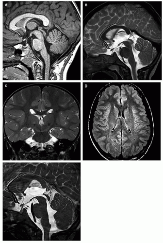
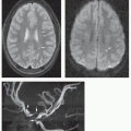
    Figure 8-4 MRI for hydrocephalus. Normal images. A. Midline sagittal T1, thin section (1.5 mm) image shows the commissures, septum pellucidum, and fornix. Note the anterior recesses of the third ventricle (chiasmatic recess in front of the chiasm and infundibular recess behind) and posterior recesses (pineal recess below the pineal gland and suprapineal recess above it). Also well seen are the midbrain with tectal plate and aqueduct, brainstem, fourth ventricle and vermis, cisterna magna, and basilar, interpeduncular, and suprasellar cisterns. B. Midline sagittal T2, thin (3.0 mm) image shows the same anatomical features. Note in addition the flow void through the aqueduct. C. Coronal T2-weighted image through the ventricular bodies and temporal horns and anterior hippocampi. Note the morphology of the third and lateral ventricles, especially temporal horns and hippocampal commissure. D. Axial FLAIR image is excellent for evaluation of the parenchyma, especially the periventricular white matter. E. High-definition CISS/FIESTA (1.0 mm or less) image is optimal for depiction of the third ventricle and its recesses, the aqueduct and fourth ventricle. Note the exquisite demonstration of the membrane of Liliequist (black arrows) extending from the dorsum sellae to the mammillary bodies. This sequence is not appropriate for demonstrating the flow voids.


  • Sagittal thin T2-FSE images best demonstrate the gross morphology of the midline structures and the major ventricular and cisternal CSF flow voids. The corpus callosum and septum pellucidum are stretched, and the fornix is displaced inferiorly and separated from the splenium by the distorted (verticalized) hippocampal commissure. The anterior third ventricle demonstrates rostral bulging of the lamina terminalis, along with widening of the supraoptic and infundibular recesses, and flattening or inferior convexity of the normally superiorly convex tuber cinereum. The suprapineal recess may be prominent also, but the extent varies with individual anatomy. The aqueduct is well demonstrated; its appearance varies with the location and severity of the obstruction: sometimes normal-looking, sometimes compressed, sometimes occluded with or without proximal widening, or (often) dilated. The aqueductal CSF flow void must be evaluated on this sagittal T2-FSE sequence, as it becomes more prominent when either the velocity or the amplitude is increased (103,120). Two factors may, singly or in association, increase the amplitude of the pulsatile movements of CSF in the aqueduct: one is the effacement of the pericerebral space, which redirects the arterial pulsations medially; the other is the decrease of compliance that results in an increased P2 peak of the pulse wave, as discussed previously. In contrast, a flow void is rarely seen in normal young infants with a large elastic pulsatile fontanelle and a small spine because pulsations are directed centrifugally (from the center of the brain to the periphery). Finally, sagittal T2-FSE images beautifully show the fourth ventricle, the cisterna magna, and the posterior fossa cisterns along with prominent, basilar artery-related signal voids in the prepontine cistern. Obviously, this sequence may also show a midline mass in the suprasellar, velum interpositum, quadrigeminal, fourth ventricular, or retrocerebellar regions.


  • High-definition, thin (submillimetric) sagittal steady-state T2 imaging (FIESTA/CISS) images are extremely important in the assessment of hydrocephalus, providing clear images of thin membranes in the ventricles, aqueduct, or cisterns; it is especially important for presurgical planning of a third ventriculostomy in order to determine the patency of the interpeduncular cistern. Postventriculostomy, together with sagittal T2-FSE, CISS demonstrates both the anatomic (patent ventriculostomy) and the functional (transventriculostomy flow void) results of the procedure. It may also reveal cisternal obstructing membranes that were not apparent before the ventricles were decompressed.


  • Axial FLAIR sequences show the axial ventricular morphology and the parenchyma. In hydrocephalic patients with a high ICP, the ependymal indentation of the stretched subependymal veins and bright signal in the periventricular and deep white matter suggest subependymal venous compression and lack of ISF absorption in the territory of the deep medullary tributaries. In contrast, periventricular bright FLAIR signal in chronic hydrocephalus suggests defective myelination, secondary to the particular vulnerability of the oligodendroglial precursors (118). The two should not be confused.


  • Although it is generally considered that T2-FSE sequences demonstrate the aqueductal flow void as well as phase-contrast CSF-flow imaging, several dedicated sequences have been promoted to enhance the demonstration of the CSF flow: steady-state free precession (121); spatial modulation of magnetization (SPAMM) (122); reversed fast imaging with steady-state precession (PSIF) (123); 3D sampling perfection with application-optimized contrast (3D-SPACE) (124). The most recent, a time-spatial labeling inversion pulse (Time-SLIP) technique uses spin labeling of the water molecules to show CSF oscillation (125), but their rapid diffusion outside the area of interest may somewhat blur the picture. Dynamic cardiac-gated cine phase-contrast imaging is another way of demonstrating the flow; it is very popular as it allows one to quantify the CSF motion in groups of subjects or patients (10,126). Special techniques that take advantage of the relative phase angle changes of moving spins can be used to quantify the flow of CSF through the foramina of Monro, aqueduct, foramen of Magendie, or brainstem cisterns (Fig. 8-5) (10) and have been proposed for the functional assessments of the shunts. CSF motion, however, is so dependent upon so many different factors that the reliability of the measurements in individual patients is uncertain.


  • Other techniques. Diffusion tensor imaging (DTI) analyzes the effects of microstructural changes in the parenchyma upon water motion (mean diffusivity [MD], fractional anisotropy [FA]); it may demonstrate a periventricular stretching of the white matter tracts (127). 1H MR spectroscopy can be used to identify changes in the energy metabolism (128). In arterial spin labeling (ASL) perfusion imaging, the blood-water itself is labeled by the MR sequence and serves as an intrinsic contrast medium that is used to investigate the blood flow noninvasively (89,129). Pseudocontinuous ASL (pCASL) is currently considered to be the most accurate ASL technique in children but, in newborns and young infants, the very short distance between the site of labeling in the neck and the site of imaging in the brain, in addition to the rapid heart rate of very young babies, makes its utility rather variable. Positive contrast cisternography or ventriculography have been suggested and are considered safe (130) but have failed to gain real acceptance as the trend in brain imaging is toward minimal invasiveness; its additional value is questionable given the versatility of MR. MR venography (MRV) may demonstrate an acquired or developmental venous compromise, primary or secondary to the hydrocephalus. Other sequences that may assist in the etiologic assessment are susceptibility imaging (blood residues), diffusion imaging (abscess, epidermoid cyst), or postcontrast T1 (leptomeningeal enhancement).


  • “Fast” MR scanning is a technique that uses single-shot, ultrafast imaging sequences. Just like a “quick” CT, it alleviates the need for sedation/anesthesia and is often used in our practices instead of CT because of mounting concerns regarding the use of ionizing radiation in the follow-up of hydrocephalus in children (see Chapter 1) (131). These heavily weighted T2 sequences are adequate for assessing the ventricular/cisternal size and morphology and to show the position of the shunt, but they have poor sensitivity and specificity for parenchymal changes (Fig. 8-6).

Finally, as hydrocephalus may occasionally be caused by, or associated with, a spinal disorder (tumor or malformation or syringomyelia), spinal imaging may be necessary when no intracranial cause is identified. The protocol should include sagittal T2 sequences of the whole spine; axial T2, T1 and possibly contrast-enhanced T1 sequences may be acquired though any region of interest. Use of fat suppression allows better recognition of soft tissue masses, fluid, or blood.


MRI and immature hydrocephalic brain

Imaging strategy in young infants is essentially the same as it is in more mature children, but a few technical points should be emphasized. In the absence of myelin, the T1 and T2 values of the parenchyma are
much longer than in the mature brain, which implies that, in TSE/FSE sequences, both T1-weighted and T2-weighted the TR should be much longer; for T2 images, the TE should be lengthened as well. FLAIR sequences are essentially noncontributory in the setting of hydrocephalus. The immature calvarium is quite expandable due to the fontanelles and thin bones; with the small size of the spinal canal relative to the skull, the CSF displacement by the arterial expansion at systole is directed peripherally (pulsations of the fontanelle) rather than caudally, so that there is no CSF movement and, consequently, no flow void within the aqueduct of a newborn infant (Fig. 8-7). Due to the incomplete development of the subcortical white matter, the field of the deep medullary tributaries of the subependymal veins appears more extensive across the thickness of the cerebral mantle relative to the territory of the subcortical veins, compared with adults; therefore, periventricular interstitial edema appears more extensive than in a mature brain, and there is greater diffusivity of the interstitial water. This periventricular edema may be more difficult to recognize against the background of high parenchymal water content on T1WI and T2WI images. Also, fibrotic changes in the ventricular walls allow better appreciation of a periventricular rim of low signal contrasting with the bright signal of both the CSF and the white matter on T2WI. Finally, because of the characteristics of hydrocephalus in infants, the use of CISS/FIESTA to demonstrate arachnoid septations, or susceptibility-weighted sequences to demonstrate blood residue is particularly useful in the assessment of the hydrocephalic brain in this age group.


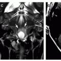
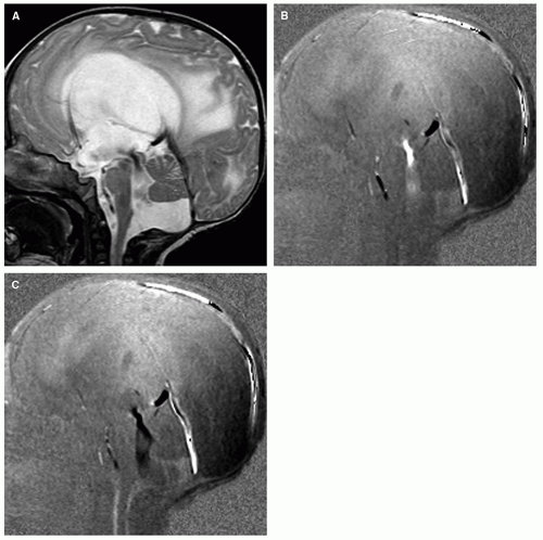
Figure 8-5 Evaluation of the CSF kinetics. A. Midline sagittal T2-weighted image. Severe hydrocephalus with a prominent flow void in the aqueduct. B. Cine phase-contrast imaging, systolic phase (compare with the flow in the straight sinus). Superior to inferior flow through the aqueduct, similar to the flow void in (A), is shown by hyperintense signal. C. Cine phase-contrast imaging, diastolic phase, shows inferior to superior flow as hypointense signal.


MRI and fetal hydrocephalic brain

Brain MRI began to be performed in the fetus as soon as MRI became a routine clinical diagnostic technique, and from the beginning, “ventriculomegaly” was a major indication (132,133). After nearly 25 years of this technique, no deleterious effects for the fetus have been reported at 1.5 T (132,133). The study always is done after a preliminary evaluation by ultrasonography. Although the volume of the fetal brain (50-60 mL about midgestation) is small relative to the volume of the maternal abdomen, the distance between the fetal brain and the abdominal surface coil is long, the fetus is quite mobile and the respiration of the mother together with the vascular and visceral motion causes artifacts, and fetal brain imaging quickly became an extremely valuable technique (134,135). The mother is placed in a supine position in the magnet (or lateral decubitus if needed). An abdominal phased array
coil is placed in such a way that the fetal head is in the center of the field explored by the coil. Each sequence serves as the localizing scout for the next one. Slices should be 2 to 3 mm thick and cover the whole fetal brain in the three orthogonal (sagittal, coronal, and axial) planes.


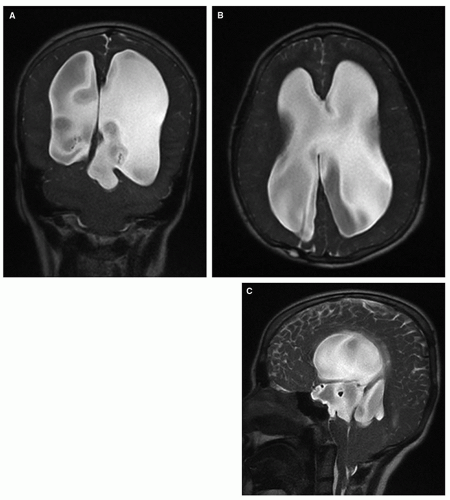
Figure 8-6 “Fast” MR imaging sequences. To avoid exposing young children to ionizing radiation, fast MR sequences (A-C) have been devised; these provide reasonably good images of the ventricles in seconds, in uncooperative hydrocephalic children but parenchymal evaluation by these is very limited.

The main technique for assessing the fetal brain is T2WI imaging using a single shot fast spin-echo (SSFSE) sequence, which shows parenchymal and ventricular anatomy nicely. Contrast in the fetal brain depends mostly on the cellular density, as gray and white matters have a similarly high content of water (90%). Additional T1WI sequences are used to show fat, calcification, and acute or subacute blood. Magnetic susceptibility sequences demonstrate blood or blood residues. Diffusion imaging is not of much use in the evaluation of fetal hydrocephalus other than in the context of an acute event. Diffusion-weighted imaging also helps to distinguish tissue types. MR spectroscopy and DTI are not used in the standard clinical routine, and no contrast is typically administered in pregnant women (134,135,136). The anatomy of the brain, the appearance of the parenchyma, and, especially, the progression of gyration must be correlated with the age, in weeks, of the fetus.


CT Scanning

Together with MR imaging, CT remains a commonly used modality in many institutions to evaluate hydrocephalus. CT is faster and very simple, and data are easily reformatted in multiple planes on modern scanners (Fig. 8-8). Periventricular interstitial edema is readily apparent on CT, and calcification may appear better on CT than on MR.
Mass effects responsible for the hydrocephalus are easily recognized, and contrast enhancement helps in the characterization of the pathology. However, the diagnostic yield and the versatility of CT are much less that those of MR, and CT can expose children to significant doses of ionizing radiation (see Chapter 1). Therefore, in our practices, initial evaluation of the brain is based on MR, as it is fast, is accurate, gives off no ionizing radiation, and is equally (or more) sensitive to small narrowings in the CSF pathways. If CT is to be used for follow-up, the implementation of a low-dose technique actually limits evaluation mostly to assessment of ventricular size (Chapter 1).


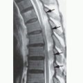
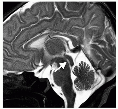
Figure 8-7 Midline sagittal T2-FSE in a neonate. Because of the large, open fontanelles, the parenchymal systolic CSF pulsations are directed outward; as a result, no flow void is seen within the aqueduct (white arrow, compare with Fig. 8-4B).


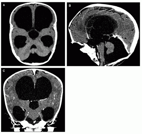
Figure 8-8 3D-CT imaging. Nice demonstration of the ventricular and cisternal anatomy in the axial (A), sagittal (B), and coronal (C) planes. Although not as sensitive and versatile as MR, modern CT is a fast and efficient diagnostic tool, and it is commonly used for screening or follow-up. Its main disadvantage is the radiation dose delivered, especially concerning in children. In most instances, a very low-dose CT (see Chapter 1) is used to check ventricular size.


Ultrasonography

Head ultrasound is the modality of choice for the screening of fetuses, neonates, and infants suspected of having hydrocephalus. It is noninvasive, can be performed at the bedside, and gives a good evaluation of the ventricular size and morphology. It is also sensitive to (if not specific for) parenchymal abnormalities and, when Doppler is used, can give an efficient functional assessment of the vasculature. Its use, however, is limited to the first months of life when the fontanelles are open, and it does not give nearly as comprehensive a presurgical assessment of hydrocephalus as MRI. Monitoring of ICP is an important factor in the differentiation of hydrocephalus from atrophy; therefore, it is important to know that innovative approaches with transfontanelle
sonography may allow assessment of ICP noninvasively. Taylor and Madsen (137) studied the hemodynamic response to fontanelle compression in infants with ventriculomegaly, and used Doppler sonography to determine resistive indices in the anterior and middle cerebral arteries in premature neonates who had suffered intracranial hemorrhage. Baseline resistive indices without fontanelle compression did not correlate with ICP. However, a significant correlation was found between the changes in resistive index during fontanelle compression in children with elevated ICP, with the maximum change in resistive index significantly higher in infants who subsequently required shunting than in infants who did not (p = 0.001) (137). Thus, the need for shunt placement may be determined by assessment of resistive indices with and without fontanelle compression.


Imaging Features of Hydrocephalus

No single feature, and especially not the ventricular dilation alone, allows the radiologist to safely make the diagnosis of hydrocephalus. The three most important features are (a) the dilatation of the anterior recesses of the third ventricle, (b) the downward bulging of the tuber cinereum, and (c) the effacement of the CSF spaces over the cerebral convexity (Table 8-3).


The Ventricular System

In children, the characteristic imaging triad of hydrocephalus includes (a) ventricular rounding and dilatation, (b) effacement of the pericerebral spaces over the convexity, and (c) macrocephaly. Normal lateral ventricles in children are typically narrow, encroached upon by the heads of the caudates, the thalami, the hippocampi, the calcarine sulci (when the occipital horn is apparent), and the collateral sulci (Fig. 8-4). In hydrocephalus, the lateral ventricles are large (Fig. 8-9) with a rounded appearance on the axial and, especially, the coronal images, notably at the level of the temporal horns (Fig. 8-9C-E). Several features allow the differentiation between hydrocephalus and ex vacuo ventriculomegaly in children (Fig. 8-10). For the frontal horns, bodies, and atria, widening and rounding are not really specific, as they can be similar in atrophy. The appearance of the temporal horns, however, is quite specific: in hydrocephalus, the horn is rounded with the choroid fissure becoming enlarged and the hippocampus being compressed and displaced inferomedially (138) (Fig. 8-9C and D), whereas in the atrophic brain, the temporal horns dilate less than the bodies of the lateral ventricles, the roof and the floor of the horn remain roughly parallel, and the hippocampus is not displaced (Fig. 8-10) (139). Note that the presence of temporal horn enlargement is less reliable in children with significant temporal lobe atrophy, such as those with Down syndrome and that the sylvian fissures should always be studied to assess the degree of temporal lobe atrophy before enlargement of the temporal horns is used to make a diagnosis of hydrocephalus. If the sylvian fissures are enlarged, or other evidence of temporal lobe atrophy is present, enlarged temporal horns are not a reliable sign of hydrocephalus. Several indices have been devised to identify hydrocephalus: the ventricular index (ratio of the ventricular diameter at the frontal horns to the diameter of the brain at the same level [Fig. 8-11A]), the ventricular angle (Fig. 8-11C) (the angle between the anterior or superior margins of the frontal horns at the level of the foramina of Monro is diminished by concentric enlargement of the frontal horns) (140), and the concentric enlargement of the frontal horns producing an appearance of “Mickey Mouse ears” on axial scans (this enlargement of the frontal horns also can be quantified by measuring the so-called frontal horn radius [Fig. 8-11D], the widest diameter of the frontal horns taken at a 90° angle to their long axis (140)). We found that the largest atrial transverse diameter of the largest ventricle, measured in the coronal plane, is the most appropriate for follow-up studies as hydrocephalus in children tends to affect the posterior parts of the hemispheres more than their anterior parts. Finally, a last point must be emphasized: the ventricular rounding (without an obvious ventricular dilation) may be the single feature identified in a “hyperacutehydrocephalus (within hours) associated with a brain swelling, because the swelling fills the pericerebral spaces and does not allow the ventricles to expand, even though they have become entrapped by the compression of the midbrain. A tamponade effect results as CSF secretion persists and can be relieved by ventricular drainage only. Such a “hyperacute” process may be observed in cases of traumatic malignant brain swelling or when a ventricular shunt becomes suddenly blocked in a treated hydrocephalus.


Table 8-3 Imaging Characteristics of Hydrocephalus (Given in Order of Utility)


  1. Enlargement of the anterior or posterior recesses of the third ventricle


  2. Downward convexity of the floor of the third ventricle (results in decreased mamillopontine distance)


  3. Effacement of the arachnoid spaces of the convexity (including the sylvian cisterns)


  4. Commensurate dilatation of the temporal horn with the lateral ventricles and rounding


  5. Atrial dilatation


The first three are the most useful signs. Although many measurements can be derived from these signs (e.g., spleniochiasmal distance, third ventricular splenial distance, mamillocommissural distance, these measurements are rarely necessary.


Dilation and tension of the lateral ventricles modifies the appearance of the midline. As the lateral ventricles are more dilated than the third ventricle, the latter is pushed downward. The corpus callosum is stretched, thinned, arched upward, more than the columns of the fornix, so that the distance between them increases (Fig. 8-9A): the septum pellucidum is stretched and may even be torn (Fig. 8-9B). The part of the fornix that forms the hippocampal commissure (the psalterium), normally transverse, becomes verticalized on each side of the midline (Fig. 8-9D, compare with Fig. 8-4C), so that the posterior part of the fornix seems to be detached from the undersurface of the splenium on the midline sagittal cut (Fig. 8-9A, compare with Fig. 8-4A).

In rare cases of chronic severe obstructive hydrocephalus (usually AS), the ventricular anatomy may be compounded by a ventricular diverticulum, inferior expansion of the medial wall of the ventricular atrium behind the thalamus, into the velum interpositum, quadrigeminal, and supracerebellar cisterns (Fig. 8-12) (141,142). This is because the inferomedial portion of the atrial wall (hippocampal commissure) is the thinnest and has the largest surface of the ventricular system and has, therefore, the highest wall tension. If not treated, these atrial diverticula can compress the midbrain with the risk of significant neurological complications; they can be mistaken for arachnoid cysts in the region of the quadrigeminal cistern (Fig. 8-12C). Coronal images are very helpful in the evaluation of these patients, demonstrating the continuity of the trigone of the lateral ventricle with the diverticulum (Fig. 8-12B).

The third ventricle is normally slit-like, mildly wider anteriorly (hypothalamic portion) than posteriorly (thalamic portion). It is enlarged in both atrophy and hydrocephalus, but in hydrocephalus, it commonly develops rounded recesses: anteriorly, the supraoptic and infundibular recesses and posteriorly, the suprapineal recess. On sagittal images (Figs. 8-9B and 8-13A), the anterior wall of the third ventricle (lamina terminalis), normally straight, becomes convex anteriorly and the tuber cinereum, usually convex upward, becomes straightened

or convex downward. This may compromise the hypothalamic nuclei and fascicles in the infundibulum and diminish the flow within the hypothalamic-pituitary portal venous system along the stalk, resulting in hypothalamic-pituitary dysfunction. When the aqueduct or the lumen or the outlets of the fourth ventricle are occluded, the tuber cinereum is characteristically hugely dilated and bulges into the interpeduncular cistern (Figs. 8-9A and B and 8-13A) sometimes wrapping the head of the basilar artery. The pulsating dilated anterior third ventricle may be so huge as to erode the dorsum sellae (Figs. 8-9A and 8-13A). When hydrocephalus is severe and chronic, the floor of the third ventricle may even rupture spontaneously, creating an internal drainage pathway that allows CSF to escape from the ventricular system into the subarachnoid space (143). The disproportionate enlargement of the recesses of the third ventricle results from the relatively small resistance to expansion provided by the thin hypothalamus and the cisterns that surround the walls of the recesses. In contrast, the thalami, which form the walls of the posterior third ventricle, provide a great deal more resistance to expansion. It is important to note that even with a significant ventricular enlargement the lateral walls of the third ventricle (i.e., the thalami) remain parallel to each other, but the massa intermedia becomes elongated and thinner (Fig. 8-9E).

On axial images, the dilated anterior recesses of the third ventricle are best detected by noting that the third ventricle is larger at the level of the optic chiasm (Fig. 8-13B) than at the level of the thalami: a rounded, circular posterior third ventricular lumen would indicate the presence of a cyst, usually a suprasellar cyst with superior expansion (Fig. 8-14). The normal suprapineal recess may be small or prominent depending on the individual anatomy; a dilated suprapineal recess of the third ventricle may sometimes expand into the posterior incisural space, displacing the pineal gland inferiorly and, occasionally, elevating the vein of Galen. Further posterior enlargement of the pineal recess may compress the tectum from the posterior direction, resulting in thinning of the tectum and narrowing of the aqueduct (144). As a rule, the anterior (supraoptic and infundibular) recesses seem to enlarge earlier and to a greater degree than the posterior (suprapineal) recess. Caudally, the dilatation may involve the proximal aqueduct with consequent shortening of the tectum in the rostral-caudal direction (144) (Fig. 8-13A). The short, thick tectum should not be mistaken for a neoplasm; isointensity with normal brain tissue on T2/FLAIR images and the absence of contrast enhancement exclude a tumor.

Only gold members can continue reading. Log In or Register to continue

Stay updated, free articles. Join our Telegram channel

Nov 11, 2018 | Posted by in NEUROLOGICAL IMAGING | Comments Off on Hydrocephalus

Full access? Get Clinical Tree

Get Clinical Tree app for offline access